SQSTM1/p62 Mouse Monoclonal Antibody


-
-
- 20μL
- ¥620
- 1-3个工作日
-
- 50μL
- ¥1250
- 1-3个工作日
-
- 100μL
- ¥2200
- 1-3个工作日
Product Details | Host Species: Mouse | Reactivity: Human,Mouse,Rat | Molecular Wt: 48 kDa | |
Clonality: Monoclonal | Isotype: IgG1 | Concentration: 1mg/ml | ||
Other Names: P62; P62/SQSTM1; sequestosome 1; SQSTM1; Ubiquitin binding protein p62 | ||||
Formulation: Liquid in PBS containing 50% glycerol, 0.5% BSA and 0.02% sodium azide. | ||||
Purification: Affinity-chromatography | ||||
Storage: Store at -20°C. Stable for one year after shipment. Aliquoting is unnecessary for -20°C storage. | ||||
Applications | WB 1:500-1:2000 | |||
Immunogen Information | Gene Name: SQSTM1 | Protein Name: Sequestosome-1 | ||
Gene ID: 8878 (Human) | SwissPro: Q13501 (Human) | |||
Subcellular Location: Cytoplasmic vesicle, autophagosome. Preautophagosomal structure. Cytoplasm, cytosol. Nucleus, PML body. Late endosome. Lysosome. Nucleus. Endoplasmic reticulum. Cytoplasm, myofibril, sarcomere. | ||||
Immunogen: Recombinant fragment of human SQSTM1/p62. AA range: 232-356. | ||||
Specificity: SQSTM1/p62 Monoclonal Antibody detects endogenous levels of SQSTM1/p62 protein. | ||||
| Product images | |
|
|
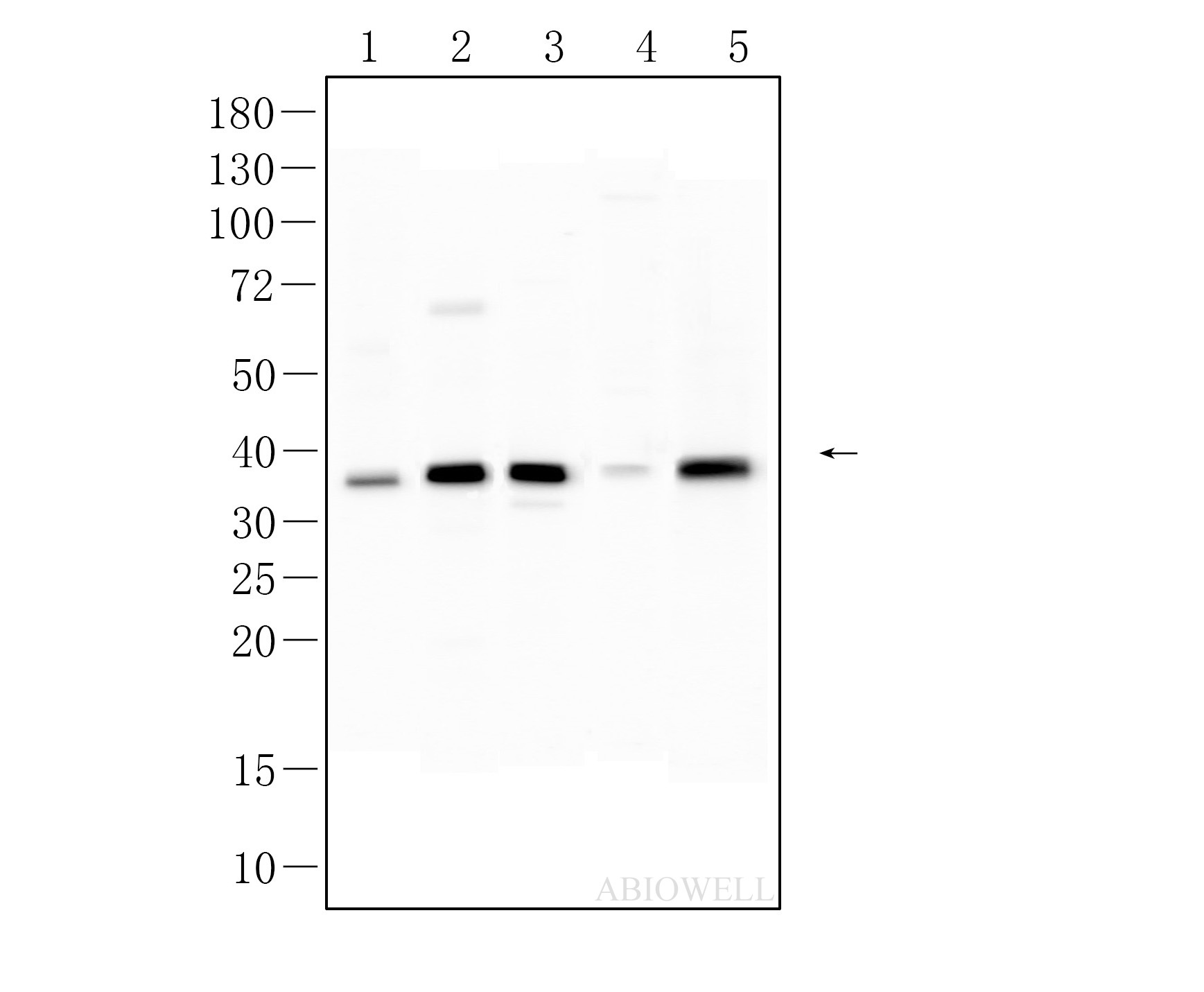
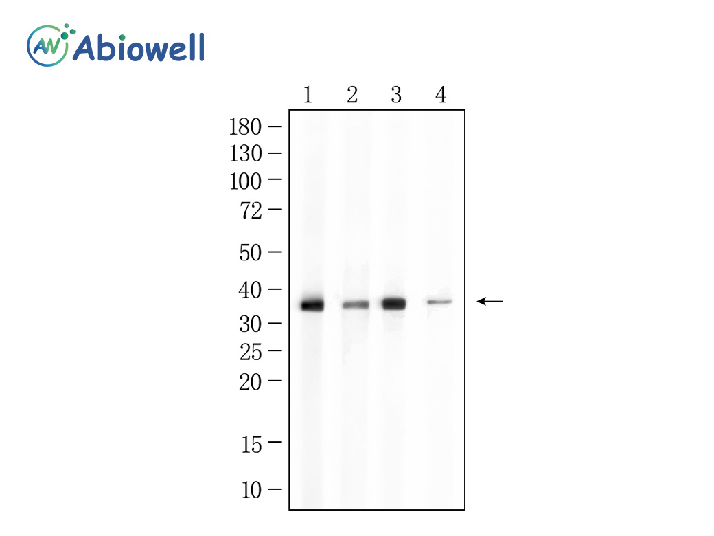
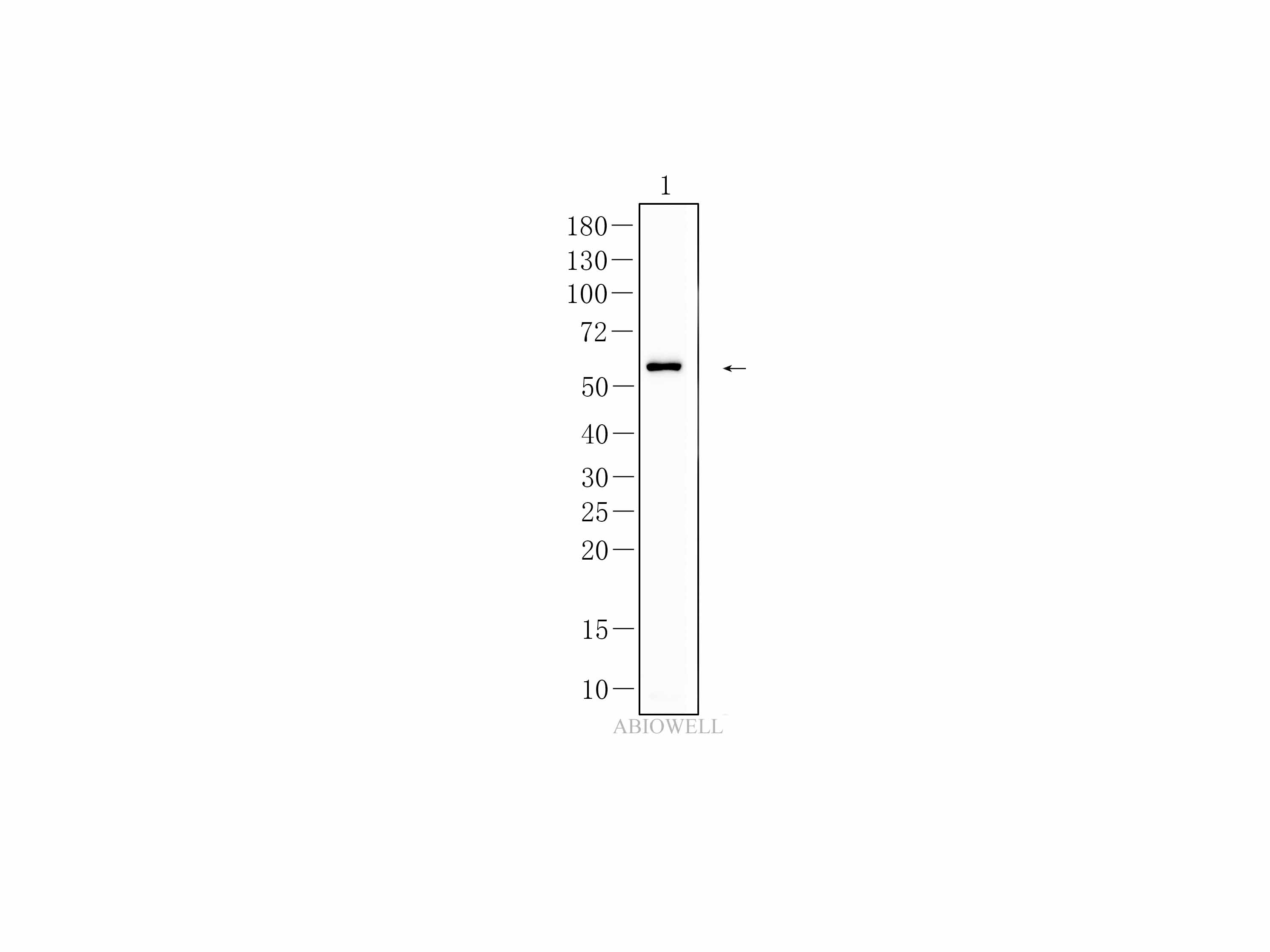
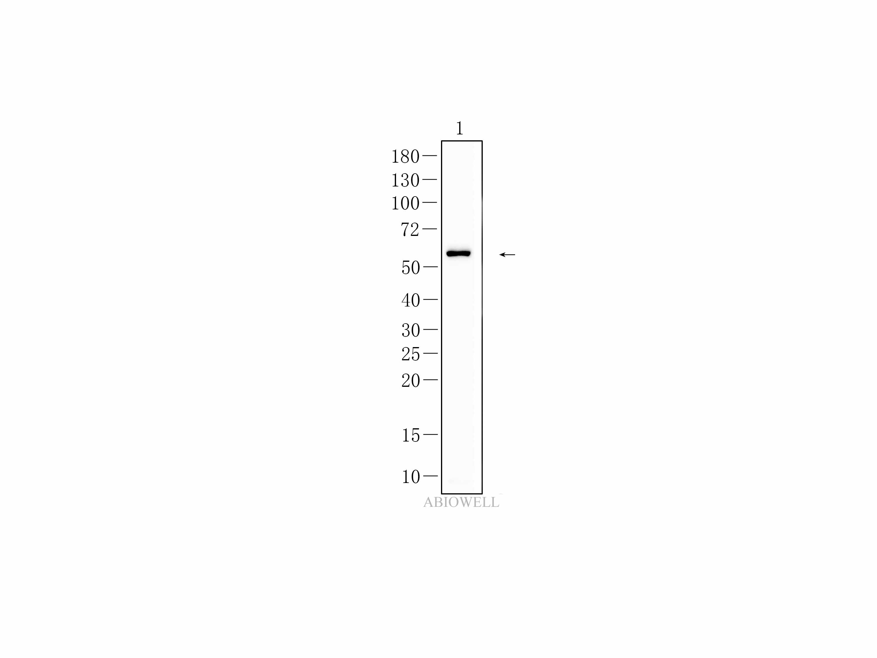
Fig : Western blot analysis of SQSTM1 on different lysates. Proteins were transferred to a NC membrane and blocked with 5% NF-Milk in TBST for 90 minutes at room temperature. The primary antibody (AWA00001, 1/1000) was used in TBST at room temperature for 90 minutes. Goat Anti-Mouse IgG - HRP Secondary Antibody (AWS0001) at 1:5,000 dilution was used for 1 hour at room temperature. Positive control: Lane 1: HEK293 cell Lane 2: NIH3T3 cell Lane 3: Hela cell |
|
|
Fig: Fluorescence immunohistochemical analysis of Mouse-colon tissue (Formalin / PFA-fixed paraffin-embedded sections) with mouse anti-SQSTM1/p62 antibody (AWA00001) at 1/100 dilution. The immunostaining was performed with the TSA Immuno-staining Kit (ABIOWELL, AWI0689). The section was pre-treated using heat mediated antigen retrieval with EDTA buffer (pH 9.0) for 20 minutes. The tissues were blocked in 5% BSA for 60 minutes at 37℃, washed with ddH2O and PBS, and then probed with the primary antibody (AWA00001) at 1/100 dilution for 1 hour at room temperature. The detection was performed using an HRP conjugated compact polymer system followed by a separate fluorescent tyramide signal amplification system (red). DAPI (blue, AWC0291) was used as a nuclear counter stain. Image acquisition was performed with Slide Scanner. |
|
|
Fig : Immunohistochemical analysis of paraffin-embedded Mouse-colon tissue with mouse anti-SQSTM1/p62 antibody (AWA00001) at 1/100 dilution. The section was pre-treated using heat mediated antigen retrieval with Sodium citrate buffer (pH 6.0) for 20 minutes. The tissues were blocked in 3% H2O2 for 15 minutes at room temperature, washed with ddH2O and PBS, and then probed with the primary antibody (AWA00001) at 1/100 dilution for 1 hour at room temperature. The detection was performed using an HRP conjugated compact polymer system(ABIOWELL, AWI0629). DAB was used as the chromogen. Tissues were counterstained with hematoxylin and mounted with DPX. |
|
|
Fig: Fluorescence immunohistochemical analysis of Mouse-cortex tissue (Formalin / PFA-fixed paraffin-embedded sections) with mouse anti-SQSTM1/p62 antibody (AWA00001) at 1/100 dilution. The immunostaining was performed with the TSA Immuno-staining Kit (ABIOWELL, AWI0689). The section was pre-treated using heat mediated antigen retrieval with EDTA buffer (pH 9.0) for 20 minutes. The tissues were blocked in 5% BSA for 60 minutes at 37℃, washed with ddH2O and PBS, and then probed with the primary antibody (AWA00001) at 1/100 dilution for 1 hour at room temperature. The detection was performed using an HRP conjugated compact polymer system followed by a separate fluorescent tyramide signal amplification system (red). DAPI (blue, AWC0291) was used as a nuclear counter stain. Image acquisition was performed with Slide Scanner. |
|
|
Fig: Fluorescence immunohistochemical analysis of Rat-colon tissue (Formalin / PFA-fixed paraffin-embedded sections) with mouse anti-SQSTM1/p62 antibody (AWA00001) at 1/100 dilution. The immunostaining was performed with the TSA Immuno-staining Kit (ABIOWELL, AWI0689). The section was pre-treated using heat mediated antigen retrieval with EDTA buffer (pH 9.0) for 20 minutes. The tissues were blocked in 5% BSA for 60 minutes at 37℃, washed with ddH2O and PBS, and then probed with the primary antibody (AWA00001) at 1/100 dilution for 1 hour at room temperature. The detection was performed using an HRP conjugated compact polymer system followed by a separate fluorescent tyramide signal amplification system (red). DAPI (blue, AWC0291) was used as a nuclear counter stain. Image acquisition was performed with Slide Scanner. |
|
|
Fig: Fluorescence immunohistochemical analysis of Mouse-hippocampus tissue (Formalin / PFA-fixed paraffin-embedded sections) with mouse anti-SQSTM1/p62 antibody (AWA00001) at 1/100 dilution. The immunostaining was performed with the TSA Immuno-staining Kit (ABIOWELL, AWI0689). The section was pre-treated using heat mediated antigen retrieval with EDTA buffer (pH 9.0) for 20 minutes. The tissues were blocked in 5% BSA for 60 minutes at 37℃, washed with ddH2O and PBS, and then probed with the primary antibody (AWA00001) at 1/100 dilution for 1 hour at room temperature. The detection was performed using an HRP conjugated compact polymer system followed by a separate fluorescent tyramide signal amplification system (red). DAPI (blue, AWC0291) was used as a nuclear counter stain. Image acquisition was performed with Slide Scanner. |
|
|
Fig : Immunohistochemical analysis of paraffin-embedded Mouse-brain tissue with mouse anti-SQSTM1/p62 antibody (AWA00001) at 1/100 dilution. The section was pre-treated using heat mediated antigen retrieval with Sodium citrate buffer (pH 6.0) for 20 minutes. The tissues were blocked in 3% H2O2 for 15 minutes at room temperature, washed with ddH2O and PBS, and then probed with the primary antibody (AWA00001) at 1/100 dilution for 1 hour at room temperature. The detection was performed using an HRP conjugated compact polymer system(ABIOWELL, AWI0629). DAB was used as the chromogen. Tissues were counterstained with hematoxylin and mounted with DPX. |
|
|
Fig : Immunohistochemical analysis of paraffin-embedded Mouse-kidney tissue with mouse anti-SQSTM1/p62 antibody (AWA00001) at 1/100 dilution. The section was pre-treated using heat mediated antigen retrieval with Sodium citrate buffer (pH 6.0) for 20 minutes. The tissues were blocked in 3% H2O2 for 15 minutes at room temperature, washed with ddH2O and PBS, and then probed with the primary antibody (AWA00001) at 1/100 dilution for 1 hour at room temperature. The detection was performed using an HRP conjugated compact polymer system(ABIOWELL, AWI0629). DAB was used as the chromogen. Tissues were counterstained with hematoxylin and mounted with DPX. |
|
|
Fig : Immunohistochemical analysis of paraffin-embedded Rat-brain tissue with mouse anti-SQSTM1/p62 antibody (AWA00001) at 1/100 dilution. The section was pre-treated using heat mediated antigen retrieval with Sodium citrate buffer (pH 6.0) for 20 minutes. The tissues were blocked in 3% H2O2 for 15 minutes at room temperature, washed with ddH2O and PBS, and then probed with the primary antibody (AWA00001) at 1/100 dilution for 1 hour at room temperature. The detection was performed using an HRP conjugated compact polymer system(ABIOWELL, AWI0629). DAB was used as the chromogen. Tissues were counterstained with hematoxylin and mounted with DPX. |
|
|
Fig: Fluorescence immunohistochemical analysis of Rat-cortex tissue (Formalin / PFA-fixed paraffin-embedded sections) with mouse anti-SQSTM1/p62 antibody (AWA00001) at 1/100 dilution. The immunostaining was performed with the TSA Immuno-staining Kit (ABIOWELL, AWI0689). The section was pre-treated using heat mediated antigen retrieval with EDTA buffer (pH 9.0) for 20 minutes. The tissues were blocked in 5% BSA for 60 minutes at 37℃, washed with ddH2O and PBS, and then probed with the primary antibody (AWA00001) at 1/100 dilution for 1 hour at room temperature. The detection was performed using an HRP conjugated compact polymer system followed by a separate fluorescent tyramide signal amplification system (red). DAPI (blue, AWC0291) was used as a nuclear counter stain. Image acquisition was performed with Slide Scanner. |
|
|
Fig : Immunohistochemical analysis of paraffin-embedded Rat-kidney tissue with mouse anti-SQSTM1/p62 antibody (AWA00001) at 1/100 dilution. The section was pre-treated using heat mediated antigen retrieval with Sodium citrate buffer (pH 6.0) for 20 minutes. The tissues were blocked in 3% H2O2 for 15 minutes at room temperature, washed with ddH2O and PBS, and then probed with the primary antibody (AWA00001) at 1/100 dilution for 1 hour at room temperature. The detection was performed using an HRP conjugated compact polymer system(ABIOWELL, AWI0629). DAB was used as the chromogen. Tissues were counterstained with hematoxylin and mounted with DPX. |
|
|
Fig : Immunohistochemical analysis of paraffin-embedded Rat-colon tissue with Mouse anti-SQSTM1/p62 antibody (AWA00001) at 1/100 dilution. The section was pre-treated using heat mediated antigen retrieval with Sodium citrate buffer (pH 6.0) for 20 minutes. The tissues were blocked in 3% H2O2 for 15 minutes at room temperature, washed with ddH2O and PBS, and then probed with the primary antibody (AWA00001) at 1/100 dilution for 1 hour at room temperature. The detection was performed using an HRP conjugated compact polymer system(ABIOWELL, AWI0629). DAB was used as the chromogen. Tissues were counterstained with hematoxylin and mounted with DPX. |
-
-
- 20μL
- ¥620
- 1-3个工作日
-
- 50μL
- ¥1250
- 1-3个工作日
-
- 100μL
- ¥2200
- 1-3个工作日
-
相关产品
-
Cdk6 Recombinant Rabbit Monoclonal Antibody
Iba1 Recombinant Rabbit Monoclonal Antibody
GAPDH Rabbit Polyclonal Antibody
GFAP Recombinant Mouse Monoclonal Antibody
Ki67 Rabbit Monoclonal Antibody
Stathmin 1 Recombinant Rabbit Monoclonal Antibody
HMGB1 Recombinant Rabbit Monoclonal Antibody
SQSTM1/p62 Mouse Monoclonal Antibody