
Goat anti-Rabbit IgG (H+L) Secondary Antibody, Alexa Fluor 488
-
-
- 50μL
- ¥260
- 现货
-
- 100μL
- ¥480
- 现货
-
- 500μL
- ¥2100
- 现货
Product Details
| Host Species: Goat | Reactivity: Rabbit | Concentration: 1 mg/mL |
Clonality: Polyclonal | Isotype: IgG | Conjugate: Alexa Fluor 488 | |
Formulation: Liquid in PBS containing 50% glycerol, 1% BSA and 0.05% PC300. | |||
Purification: Affinity-chromatography | |||
Storage: -20°C,store in dark,1 year | |||
Applications
| IF 1:100-1:1000 FCM 1:100-1:1000 | ||
Information
| Based on immunoelectrophoresis and/or ELISA, the antibody reacts with whole molecule rabbit IgG. It also reacts with the light chains of other rabbit immunoglobulins. No antibody was detected against non-immunoglobulin serum proteins. The antibody may cross-react with immunoglobulins from other species. | ||
| Product images | |
|
|
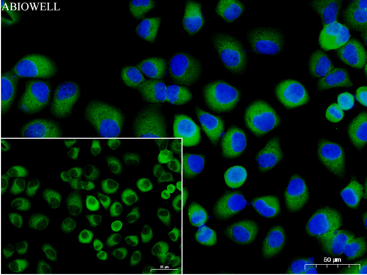
Fig: Immunocytochemistry analysis of Hela cells labeling Goat Anti-Rabbit IgG H&L (iFluor™ 488, AWS0005c) was used as the secondary antibody at 1/200 dilution for 60 minutes at 37 ℃. Nuclear DNA was labelled in blue with DAPI(AWC0291). |
|
|
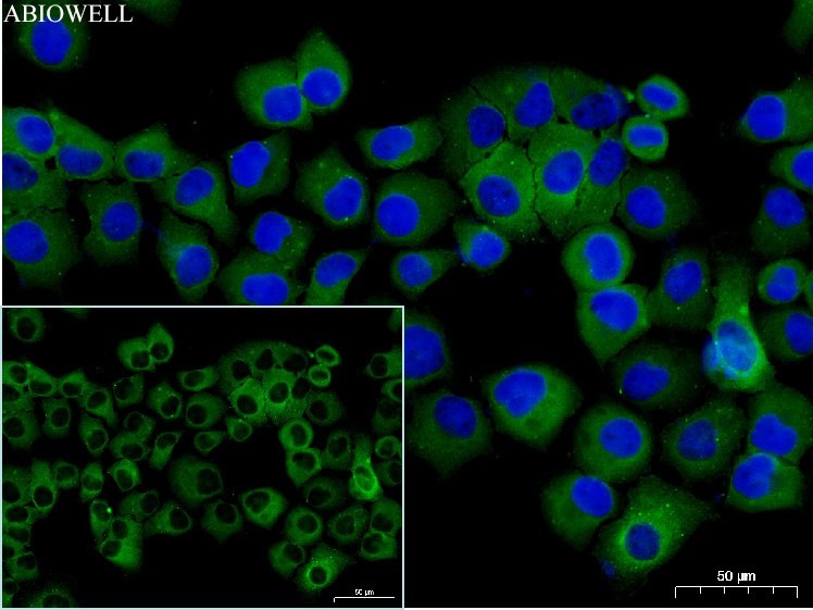
Fig: Immunocytochemistry analysis of Hela cells labeling Goat Anti-Rabbit IgG H&L (iFluor™ 488, AWS0005c) was used as the secondary antibody at 1/200 dilution for 60 minutes at 37 ℃. Nuclear DNA was labelled in blue with DAPI(AWC0291). |
|
|
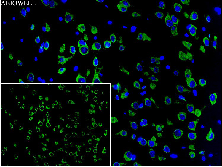
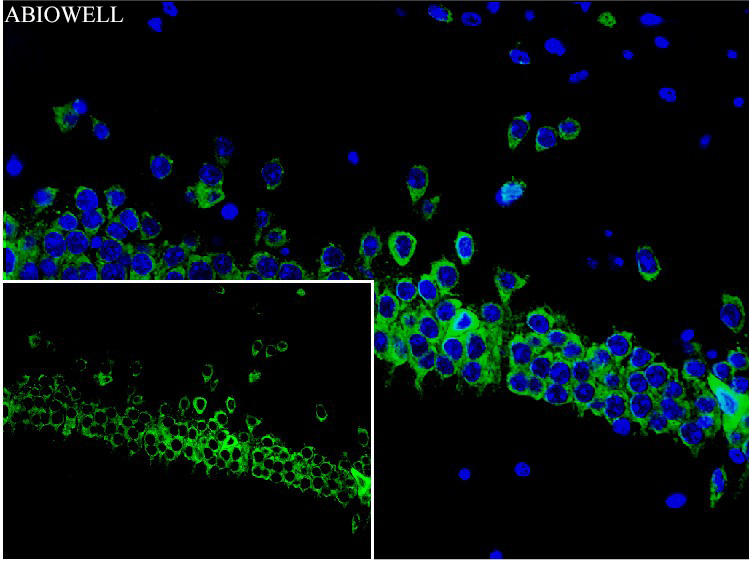
Fig: Fluorescence immunohistochemical analysis of MOUSE-brain cortex tissue (Formalin/PFA-fixed paraffin-embedded sections). Goat Anti-RABBIT IgG H&L (iFluor™ 488, AWS0005) was used as the secondary antibody at 1/200 dilution for 60 minutes at 37 ℃.DAPI (blue, AWC0291) was used as a nuclear counter stain. Image acquisition was performed with Slide Scanner. |
|
|
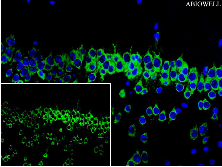
Fig: Fluorescence immunohistochemical analysis of Mouse-hippocampus tissue (Formalin/PFA-fixed paraffin-embedded sections). Goat Anti-RABBIT IgG H&L (iFluor™ 488, AWS0005) was used as the secondary antibody at 1/200 dilution for 60 minutes at 37 ℃.DAPI (blue, AWC0291) was used as a nuclear counter stain. Image acquisition was performed with Slide Scanner. |
|
|
Fig: Fluorescence immunohistochemical analysis of Mouse-hippocampus tissue (Formalin/PFA-fixed paraffin-embedded sections). Goat Anti-RABBIT IgG H&L (iFluor™ 488, AWS0005) was used as the secondary antibody at 1/200 dilution for 60 minutes at 37 ℃.DAPI (blue, AWC0291) was used as a nuclear counter stain. Image acquisition was performed with Slide Scanner. |
1. Yang, Min et al. “Lysine demethylase KDM3A alleviates hyperoxia-induced bronchopulmonary dysplasia in mice by promoting ETS1 expression.” Experimental cell research vol. 435,2 (2024): 113945. doi:10.1016/j.yexcr.2024.113945. PubMed:38286256
2. Chen, Pan et al. “ACSL4 promotes ferroptosis and M1 macrophage polarization to regulate the tumorigenesis of nasopharyngeal carcinoma.” International immunopharmacology vol. 122 (2023): 110629. doi:10.1016/j.intimp.2023.110629. PubMed:37451020
3. Li, Zengshi et al. “WNTA5-mediated miR-374a-5p regulates vascular smooth muscle cell phenotype transformation and M1 macrophage polarization impacting intracranial aneurysm progression.” Scientific reports vol. 14,1 559. 4 Jan. 2024, doi:10.1038/s41598-024-51243-z. PubMed:38177414
4. Tian, Zhenyang et al. “LncARSR promotes glioma tumor growth by mediating glycolysis through the STAT3/HK2 axis.” Cytokine vol. 180 (2024): 156663. doi:10.1016/j.cyto.2024.156663. PubMed:38815522
-
-
- 50μL
- ¥260
- 现货
-
- 100μL
- ¥480
- 现货
-
- 500μL
- ¥2100
- 现货
-
相关产品
-
Goat anti-Mouse IgG (H+L) Secondary Antibody, HRP
Goat anti-Rabbit IgG (H+L) Secondary Antibody, HRP
Goat anti-Mouse IgG (H+L) Secondary Antibody, Alexa Fluor 488
Goat anti-Mouse IgG (H+L) Secondary Antibody, Alexa Fluor 594
Goat anti-Rabbit IgG (H+L) Secondary Antibody, Alexa Fluor 488
Goat anti-Rabbit IgG (H+L) Secondary Antibody, Alexa Fluor 594
Sheep anti-Mouse IgG (H+L) Secondary Antibody, Alexa Fluor 488
Sheep anti-Mouse IgG (H+L) Secondary Antibody, Alexa Fluor 594


