
GFAP Recombinant Mouse Monoclonal Antibody
-
-
- 20μL
- ¥620
- 1-3个工作日
-
- 50μL
- ¥1250
- 1-3个工作日
-
- 100μL
- ¥2200
- 1-3个工作日
Product Details | Host Species: Mouse | Reactivity: Human,Mouse,Rat | Molecular Wt: 50 kDa | |
Clonality: Monoclonal | Isotype: IgG1 | Concentration: 1mg/ml | ||
Other Names: GFAP; Glial fibrillary acidic protein; ALXDRD; cb345; etID36982.3; FLJ42474; FLJ45472; gfapl; Intermediate filament protein | ||||
Formulation: Liquid in PBS containing 50% glycerol, 0.5% BSA and 0.02% sodium azide. | ||||
Purification: Affinity-chromatography | ||||
Storage: Store at -20°C. Stable for one year after shipment. Aliquoting is unnecessary for -20°C storage. | ||||
Applications | WB 1:500-1:2000 | |||
Immunogen Information | Gene Name: GFAP | Protein Name: Glial fibrillary acidic protein | ||
Gene ID: 2670 (Human) | SwissPro: P14136 (Human) | |||
Subcellular Location: Cytoplasm. | ||||
Immunogen: Synthetic peptide amino acids 411-422 of human GFAP. | ||||
Specificity: GFAP Monoclonal Antibody detects endogenous levels of GFAP protein. | ||||
| Product images | |
|
|
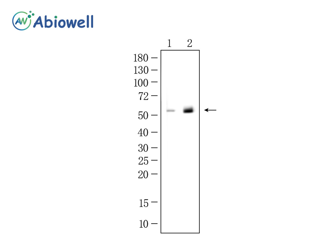
Fig : Western blot analysis of GFAP on different lysates. Proteins were transferred to a NC membrane and blocked with 5% NF-Milk in TBST for 1 hour at room temperature. The primary antibody (AWA06001, 1/1000) was used in TBST at room temperature for 2 hours. Goat Anti- Mouse IgG - HRP Secondary Antibody (AWS0001) at 1:5,000 dilution was used for 1 hour at room temperature. Positive control: Lane 1: N-2a cell Lane 2: Rat brain Predicted molecular weight:50 kDa Observed molecular weight:52 kDa Exposure time:45s |
|
|
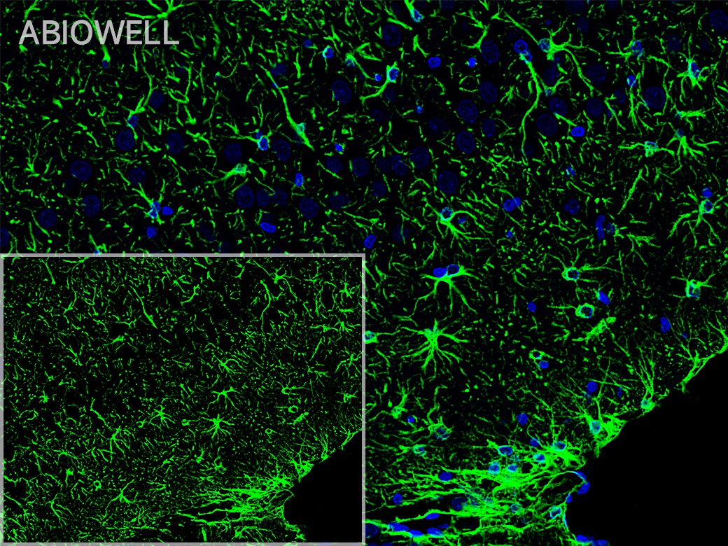
Fig: Fluorescence immunohistochemical analysis of Rat-brain tissue (Formalin/PFA-fixed paraffin-embedded sections). with Mouse anti-GFAPantibody (AWA06001) at 1/200 dilution. The immunostaining was performed with the TSA Immuno-staining Kit (ABIOWELL, I0688). The section was pre-treated using heat mediated antigen retrieval with Sodium citrate buffer (pH 6.0) for 20 minutes. The tissues were blocked in 3% H2O2 for 15 minutes at room temperature, washed with ddH2O and PBS, and then probed with the primary antibody (AWA06001) at 1/200 dilution for 2 hour at 37℃or overnignt at 4℃. The detection was performed using an HRP conjugated compact polymer system followed by a separate fluorescent tyramide signal amplification system (green). DAPI (blue, AWC0291) was used as a nuclear counter stain. Image acquisition was performed with Slide Scanner. |
|
|
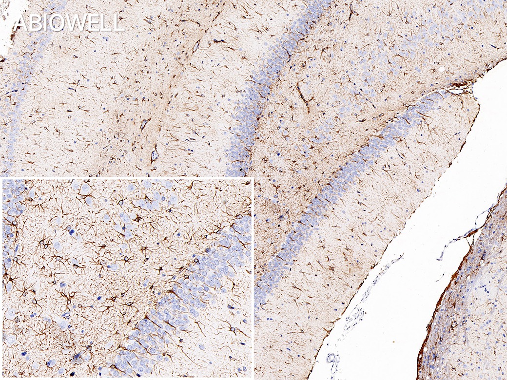
Fig : Immunohistochemical analysis of paraffin-embedded Rat-hippocampal formation tissue with Rabbit anti-GFAP antibody (AWA06001) at 1/200 dilution. The section was pre-treated using heat mediated antigen retrieval with Sodium citrate buffer (pH 6.0) for 20 minutes. The tissues were blocked in 3% H2O2 for 15 minutes at room temperature, washed with ddH2O and PBS, and then probed with the primary antibody (AWA06001) at 1/200 dilution for 2 hour at 37℃or overnignt at 4℃. The detection was performed using an HRP conjugated compact polymer system(ABIOWELL, AWI0629). DAB was used as the chromogen. Tissues were counterstained with hematoxylin and mounted with DPX. |
-
-
- 20μL
- ¥620
- 1-3个工作日
-
- 50μL
- ¥1250
- 1-3个工作日
-
- 100μL
- ¥2200
- 1-3个工作日
-
相关产品
-
Cdk6 Recombinant Rabbit Monoclonal Antibody
GAPDH Rabbit Polyclonal Antibody
GFAP Recombinant Mouse Monoclonal Antibody
Ki67 Rabbit Monoclonal Antibody
HMGB1 Recombinant Rabbit Monoclonal Antibody
SQSTM1/p62 Mouse Monoclonal Antibody
Bcl-2 Recombinant Rabbit Monoclonal Antibody
SOD2 Rabbit Polyclonal Antibody


