
COX IV Rabbit Polyclonal Antibody
-
-
- 20μL
- ¥620
- 1-3个工作日
-
- 50μL
- ¥1250
- 1-3个工作日
-
- 100μL
- ¥2200
- 1-3个工作日
Product Details | Host Species: Rabbit | Reactivity: Human,Mouse,Rat | Molecular Wt: 20 kDa | |
Clonality: Polyclonal | Isotype: IgG | Concentration: 1mg/ml | ||
Other Names: COX4I1; COX4; Cytochrome c oxidase subunit 4 isoform 1, mitochondrial; Cytochrome c oxidase polypeptide IV; Cytochrome c oxidase subunit IV isoform 1; COX IV-1 | ||||
Formulation: Liquid in PBS containing 50% glycerol, 0.5% BSA and 0.02% sodium azide. | ||||
Purification: Affinity-chromatography | ||||
Storage: Store at -20°C. Stable for one year after shipment. Aliquoting is unnecessary for -20°C storage. | ||||
Applications | WB 1:1000-1:5000 | |||
Immunogen Information | Gene Name: COX4I1 | Protein Name: Cytochrome c oxidase subunit 4 isoform 1, mitochondrial | ||
Gene ID: 1327 (Human) | SwissPro: P13073 (Human) | |||
Subcellular Location: Mitochondrion inner membrane. | ||||
Immunogen: Recombinant protein within human COX IV. AA range: 23-98. | ||||
Specificity: COX IV Polyclonal Antibody detects endogenous levels of COX IV protein. | ||||
| Product images | |
|
|
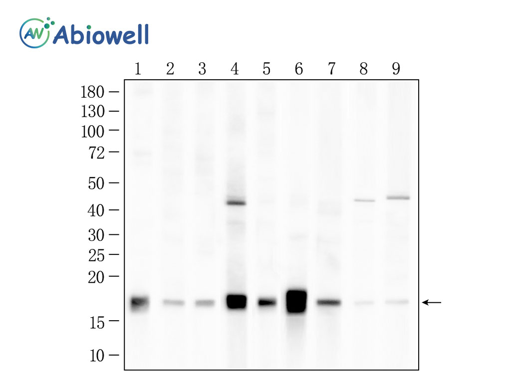
Fig : Western blot analysis of COX IV on different lysates. Proteins were transferred to a NC membrane and blocked with 5% NF-Milk in TBST for 1 hour at room temperature. The primary antibody (AWA61720, 1/1000) was used in TBST at room temperature for 2 hours. Goat Anti-Rabbit IgG - HRP Secondary Antibody (AWS0002) at 1:5,000 dilution was used for 1 hour at room temperature. Positive control: Lane 1: 5637 cell Lane 2: U87-MG cell Lane 3: SK-BR-3 cell Lane 4: HELA cell Lane 5: THP-1 cell Lane 6: HEPG2 cell Lane 7: A549 cell Lane 8: 4T1 cell Lane 9: GL261 cell Predicted molecular weight:20 kDa Observed molecular weight:17 kDa Exposure time:45seconds |
|
|
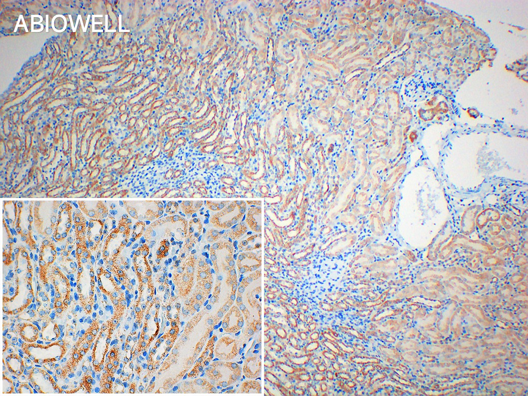
Fig : Immunohistochemical analysis of paraffin-embedded Mouse-Kidney tissue with Rabbit anti-COX Ⅳ antibody (AWA61720) at 1/200 dilution. The section was pre-treated using heat mediated antigen retrieval with Sodium citrate buffer (pH 6.0) for 20 minutes. The tissues were blocked in 3% H2O2 for 15 minutes at room temperature, washed with ddH2O and PBS, and then probed with the primary antibody (AWA61720) at 1/200 dilution for 2 hour at 37℃or overnignt at 4℃. The detection was performed using an HRP conjugated compact polymer system(ABIOWELL, AWI0629). DAB was used as the chromogen. Tissues were counterstained with hematoxylin and mounted with DPX. |
-
-
- 20μL
- ¥620
- 1-3个工作日
-
- 50μL
- ¥1250
- 1-3个工作日
-
- 100μL
- ¥2200
- 1-3个工作日
-
相关产品
-
Cdk6 Recombinant Rabbit Monoclonal Antibody
GAPDH Rabbit Polyclonal Antibody
GFAP Recombinant Mouse Monoclonal Antibody
Ki67 Rabbit Monoclonal Antibody
HMGB1 Recombinant Rabbit Monoclonal Antibody
SQSTM1/p62 Mouse Monoclonal Antibody
Bcl-2 Recombinant Rabbit Monoclonal Antibody
SOD2 Rabbit Polyclonal Antibody


