
CEBPB Rabbit Polyclonal Antibody
-
-
- 20μL
- ¥620
- 1-3个工作日
-
- 50μL
- ¥1250
- 1-3个工作日
-
- 100μL
- ¥2200
- 1-3个工作日
Product Details
| Host Species: Rabbit | Reactivity: Human,Rat | Molecular Wt: 36 kDa | |
Clonality: Polyclonal | Isotype: IgG | Concentration: 1 mg/ml | ||
Other Names: CEBPB; LAP; TCF5; PP9092; CCAAT/enhancer-binding protein beta; C/EBP beta; Liver activator protein; Nuclear factor NF-IL6; Transcription factor 5; TCF-5
| ||||
Formulation: Liquid in PBS containing 50% glycerol, 0.5% BSA and 0.02% sodium azide. | ||||
Purification: Affinity-chromatography | ||||
Storage: -20°C,1 year | ||||
Applications
| WB 1:500-1:2000
| |||
Immunogen Information | Gene Name: CEBPB | Protein Name: CCAAT/enhancer-binding protein beta | ||
Gene ID: 1051 (Human) 12608 (Mouse) 24253 (Rat)
| SwissPro: P17676 (Human) P28033 (Mouse) P21272 (Rat)
| |||
Subcellular Location: Nucleus. Cytoplasm. | ||||
Immunogen: Synthetic peptide of human CEBPB. | ||||
Specificity: CEBPB Polyclonal Antibody detects endogenous levels of EfCEBPB protein. | ||||
| Product images | |
|
|
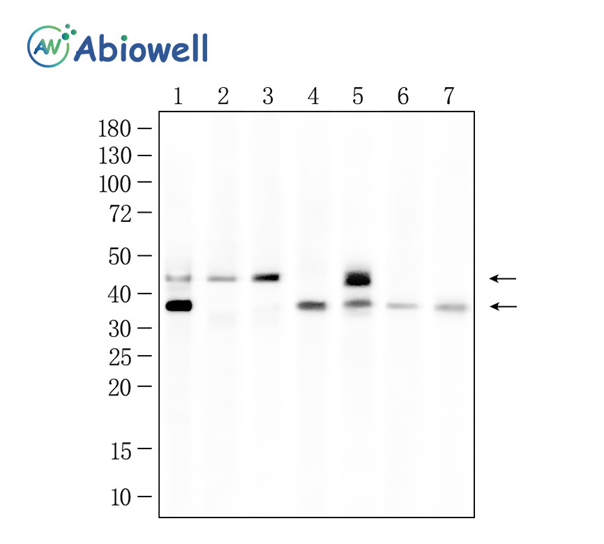
Fig : Western blot analysis of CEBPB on different lysates. Proteins were transferred to a NC membrane and blocked with 5% NF-Milk in TBST for 1 hour at room temperature. The primary antibody (AWA59024, 1/1000) was used in TBST at room temperature for 2 hours. Goat Anti- Rabbit IgG - HRP Secondary Antibody (AWS0002) at 1:5,000 dilution was used for 1 hour at room temperature. Positive control: Lane 1: MCF-7 cell Lane 2: HELA cell Lane 3: A549 cell Lane 4: SIHa cell Lane 5: HaCaT cell Lane 6: HEPG2 cell Lane 7: Rat liver Predicted molecular weight:36 kDa Observed molecular weight:36 kDa、42 kDa Exposure time:90 sec |
-
-
- 20μL
- ¥620
- 1-3个工作日
-
- 50μL
- ¥1250
- 1-3个工作日
-
- 100μL
- ¥2200
- 1-3个工作日
-
相关产品
-
Cdk6 Recombinant Rabbit Monoclonal Antibody
GAPDH Rabbit Polyclonal Antibody
GFAP Recombinant Mouse Monoclonal Antibody
Ki67 Rabbit Monoclonal Antibody
HMGB1 Recombinant Rabbit Monoclonal Antibody
SQSTM1/p62 Mouse Monoclonal Antibody
Bcl-2 Recombinant Rabbit Monoclonal Antibody
SOD2 Rabbit Polyclonal Antibody


