
MFN1 Rabbit Polyclonal Antibody
-
-
- 20μL
- ¥620
- 1-3个工作日
-
- 50μL
- ¥1250
- 1-3个工作日
-
- 100μL
- ¥2200
- 1-3个工作日
Product Details
| Host Species: Rabbit | Reactivity: Human | Molecular Wt: 84 kDa | |
Clonality: Polyclonal | Isotype: IgG | Concentration: 1 mg/ml | ||
Other Names: Fzo homolog; hfzo1; hfzo2; MFN 1; MFN1; mitofusin 1; Mitofusin 1; Mitofusin-1; Mitofusin1; MS996; Transmembrane GTPase MFN1; Mitochondrial transmembrane GTPase Fzo 1; Putative transmembrane GTPase
| ||||
Formulation: Liquid in PBS containing 50% glycerol, 0.5% BSA and 0.02% sodium azide. | ||||
Purification: Affinity-chromatography | ||||
Storage: -20°C,1 year | ||||
Applications
| WB 1:500-1:2000 | |||
Immunogen Information | Gene Name: MFN1 | Protein Name: Mitofusin-1 | ||
Gene ID: 55669 (Human) | SwissPro: Q8IWA4 (Human) | |||
Subcellular Location: Mitochondrion outer membrane. Cytoplasm. | ||||
Immunogen: Synthetic peptide of human MFN1. | ||||
Specificity: MFN1 Polyclonal Antibody detects endogenous levels of MFN1 protein. | ||||
| Product images | |
|
|
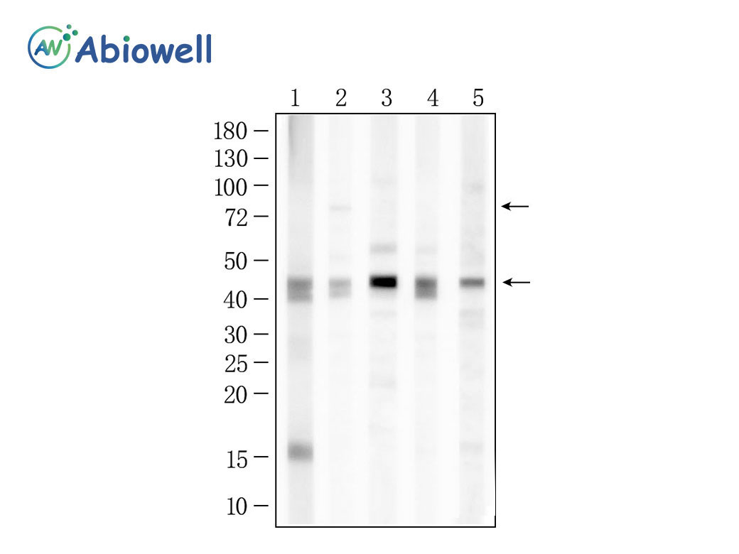
Fig : Western blot analysis of MFN1 on different lysates. Proteins were transferred to a NC membrane and blocked with 5% NF-Milk in TBST for 1 hour at room temperature. The primary antibody (AWA59015, 1/1000) was used in TBST at room temperature for 2 hours. Goat Anti-Rabbit IgG - HRP Secondary Antibody (AWS0002) at 1:5,000 dilution was used for 1 hour at room temperature. Positive control: Lane 1: T24 cell Lane 2: LN229 cell Lane 3: SW480 cell Lane 4: AGS cell Lane 5: A2780 cell Predicted molecular weight:84KD,41KD(Isoforms) Observed molecular weight:84KD ,41KD(Isoforms) Exposure time:45seconds |
-
-
- 20μL
- ¥620
- 1-3个工作日
-
- 50μL
- ¥1250
- 1-3个工作日
-
- 100μL
- ¥2200
- 1-3个工作日
-
相关产品
-
Cdk6 Recombinant Rabbit Monoclonal Antibody
GAPDH Rabbit Polyclonal Antibody
GFAP Recombinant Mouse Monoclonal Antibody
Ki67 Rabbit Monoclonal Antibody
HMGB1 Recombinant Rabbit Monoclonal Antibody
SQSTM1/p62 Mouse Monoclonal Antibody
Bcl-2 Recombinant Rabbit Monoclonal Antibody
SOD2 Rabbit Polyclonal Antibody


