
Parkin Rabbit Polyclonal Antibody
-
-
- 20μL
- ¥620
- 1-3个工作日
-
- 50μL
- ¥1250
- 1-3个工作日
-
- 100μL
- ¥2200
- 1-3个工作日
Product Details | Host Species: Rabbit | Reactivity: Human,Mouse,Rat,Chicken | Molecular Wt: 52 kDa | |
Clonality: Polyclonal | Isotype: IgG | Concentration: 1mg/ml | ||
Other Names: PARK 2; PARK2; PRKN; PRKN2; E3 ubiquitin ligase; E3 ubiquitin-protein ligase parkin; Parkinson juvenile disease protein 2; Parkinson disease protein 2; FRA6E; Park2; Parkin 2; AR JP; LPRS 2; LPRS2; PARK2/Parkin; Parkin; PDJ | ||||
Formulation: Liquid in PBS containing 50% glycerol, 0.5% BSA and 0.02% sodium azide. | ||||
Purification: Affinity-chromatography | ||||
Storage: Store at -20°C. Stable for one year after shipment. Aliquoting is unnecessary for -20°C storage. | ||||
Applications | WB 1:500-1:2000 | |||
Immunogen Information | Gene Name: PRKN | Protein Name: E3 ubiquitin-protein ligase parkin | ||
Gene ID: 5071 (Human) | SwissPro: O60260 (Human) | |||
Subcellular Location: Cytoplasm, cytosol. Nucleus. Endoplasmic reticulum. Mitochondrion. Mitochondrion outer membrane. Cell projection, neuron projection. Postsynaptic density. Presynapse. | ||||
Immunogen: The antiserum was produced against synthesized peptide derived from human Parkin. AA range: 101-150. | ||||
Specificity: Parkin Polyclonal Antibody detects endogenous levels of Parkin protein. | ||||
| Product images | |
|
|
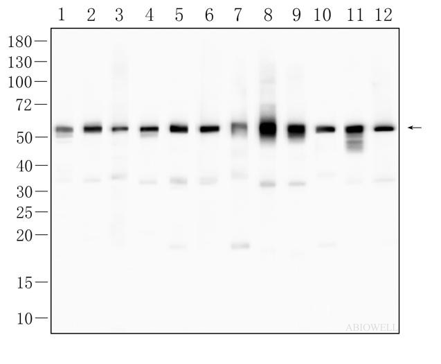
Fig : Western blot analysis of Parkin on different lysates. Proteins were transferred to a NC membrane and blocked with 5% NF-Milk in TBST for 1 hour at room temperature. The primary antibody ( AWA41194, 1/1000) was used in PBST at room temperature for 2 hours. Goat Anti-Rabbit IgG - HRP Secondary Antibody (AWS0002) at 1:5,000 dilution was used for 1 hour at room temperature. Positive control: Lane 1: Hela cell Lane 2: HUVEC cell Lane 3: Hek293T cell Lane 4: U251 cell Lane 5: GL261 cell Lane 6: SH-SY5Y cell Lane 7: HT22 cell Lane 8: N2A cell Lane 9: PC-12 cell Lane 10: NRK-49F cell Lane 11: PC-9 cell Lane 12: LLC cell Predicted molecular weight: 52 kDa Observed molecular weight: 55 kDa Exposure time: 7 seconds |
|
|
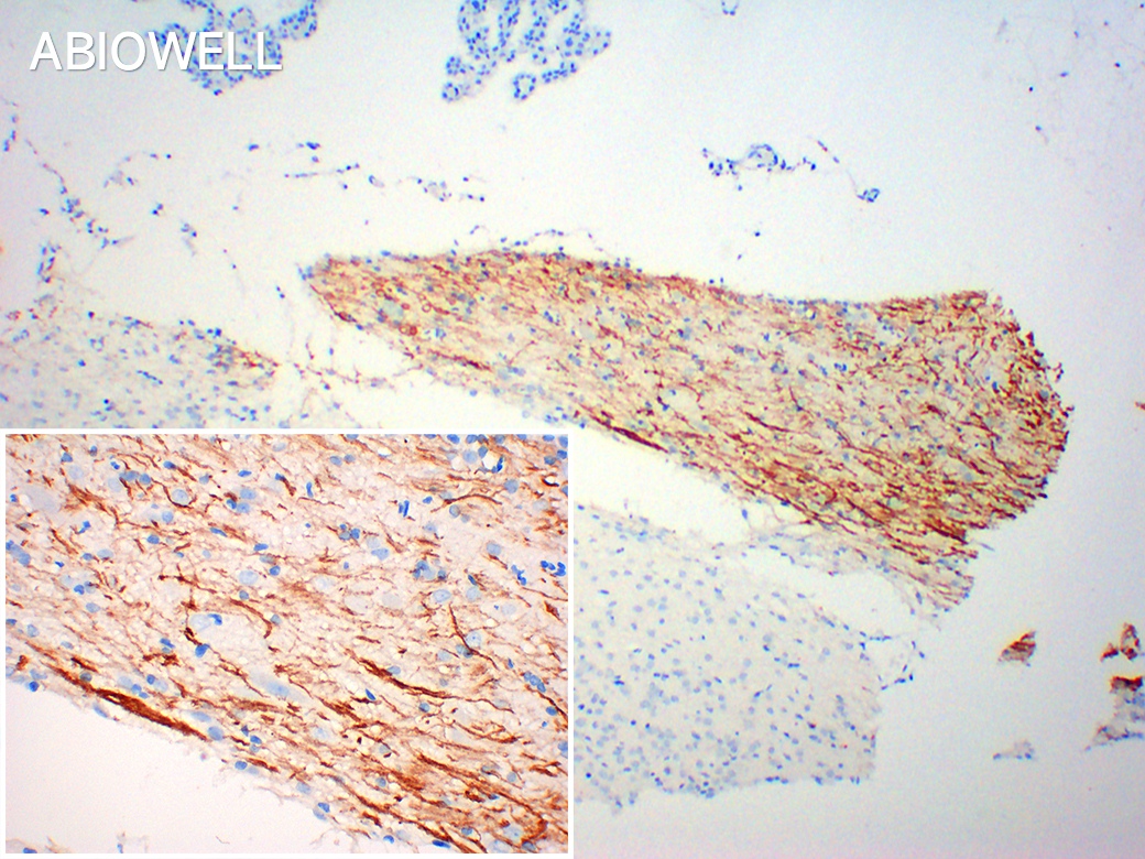
Fig : Immunohistochemical analysis of paraffin-embedded Mouse-testis tissue with Rabbit anti-Parkin antibody (AWA41194) at 1/200 dilution. The section was pre-treated using heat mediated antigen retrieval with Sodium citrate buffer (pH 6.0) for 20 minutes. The tissues were blocked in 3% H2O2 for 15 minutes at room temperature, washed with ddH2O and PBS, and then probed with the primary antibody (AWA41194) at 1/200 dilution for 2 hour at 37℃ or overnignt at 4℃. The detection was performed using an HRP conjugated compact polymer system(ABIOWELL, AWI0629). DAB was used as the chromogen. Tissues were counterstained with hematoxylin and mounted with DPX. |
-
-
- 20μL
- ¥620
- 1-3个工作日
-
- 50μL
- ¥1250
- 1-3个工作日
-
- 100μL
- ¥2200
- 1-3个工作日
-
相关产品
-
Cdk6 Recombinant Rabbit Monoclonal Antibody
GAPDH Rabbit Polyclonal Antibody
GFAP Recombinant Mouse Monoclonal Antibody
Ki67 Rabbit Monoclonal Antibody
HMGB1 Recombinant Rabbit Monoclonal Antibody
SQSTM1/p62 Mouse Monoclonal Antibody
Bcl-2 Recombinant Rabbit Monoclonal Antibody
SOD2 Rabbit Polyclonal Antibody


