
C3 Recombinant Rabbit Monoclonal Antibody
-
-
- 20μL
- ¥620
- 有库存
-
- 50μL
- ¥1250
- 有库存
-
- 100μL
- ¥2200
- 有库存
Product Details
| Host Species: Rabbit | Reactivity: Human,Mouse,Rat | Molecular Wt: 187 kDa | |
Clonality: Monoclonal | Isotype: IgG | Concentration: 1 mg/ml | ||
Other Names: ARMD9;ASP;C3;CO3_HUMAN;Complement C3;Complement factor 3;CPAMD1;HEL S 62p;Complement C3c alpha' chain fragment 2;Complement component 3
| ||||
Formulation: Liquid in PBS containing 50% glycerol, 0.5% BSA and 0.02% sodium azide. | ||||
Purification: Affinity-chromatography | ||||
Storage: -20°C,1 year | ||||
Applications
| WB 1:1000 IF 1:50-1:100 FC 1:50-1:100
| |||
Immunogen Information | Gene Name: C3 | Protein Name: Complement C3 | ||
Gene ID: 718 (Human) 12266 (Mouse) 24232 (Rat) | SwissPro: P01024 (Human) P01027 (Mouse) P01026 (Rat)
| |||
Immunogen: Synthetic peptide within Human C3 aa 1210-1248 / 1663. | ||||
Specificity: C3 Monoclonal Antibody detects endogenous levels of C3 protein. | ||||
| Product images | |
|
|
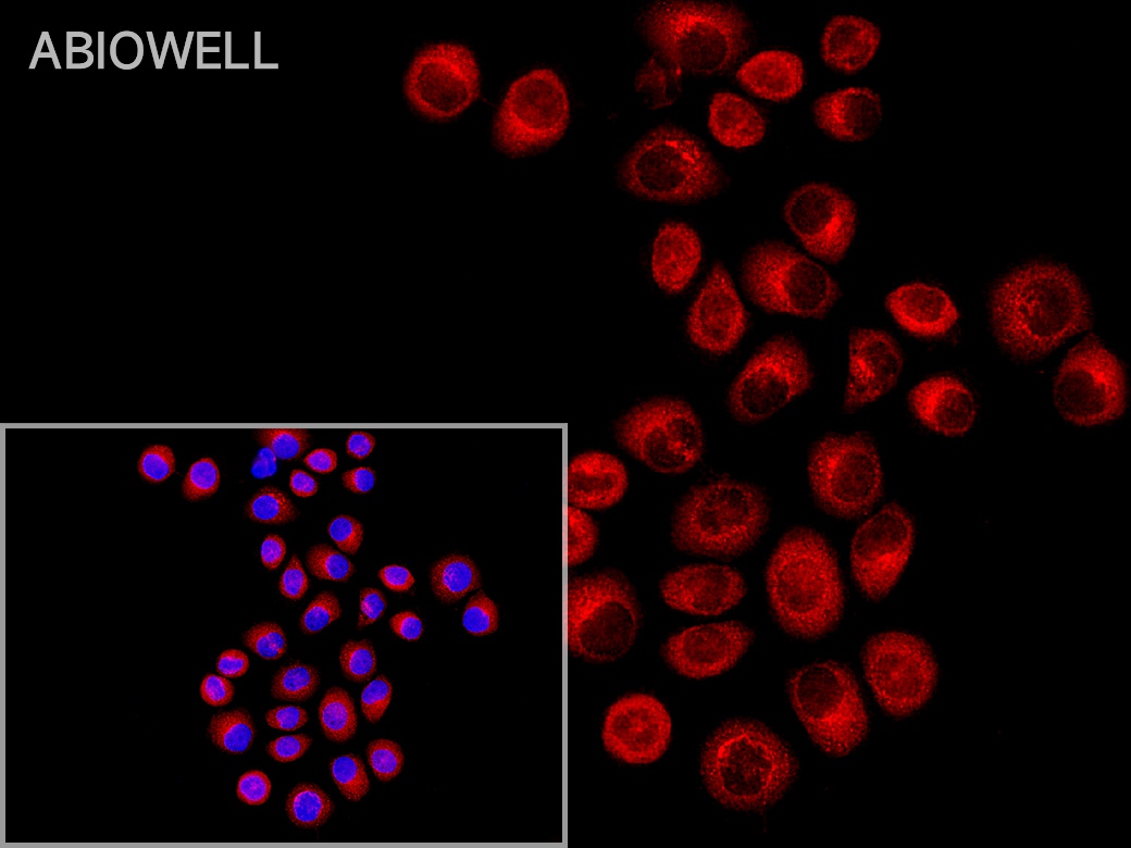
Fig: Immunocytochemistry analysis of HELA cells labeling C3 with Rabbit anti-C3 antibody (AWA12697) at 1/50 dilution(Red ). Cells were fixed in 4% paraformaldehyde for 10 minutes at 37 ℃, permeabilized with 0.03% Triton X-100 in PBS for 30 minutes, and then blocked with 5% BSA for 60 minutes at 37 ℃. Cells were then incubated with Rabbit anti-C3 antibody (AWA12697) at 1/50 dilution in 2% negative goat serum overnight at 4 ℃. Goat Anti-Rabbit IgG H&L (iFluor™ 594, AWS0006) was used as the secondary antibody at 1/200 dilution for 60 minutes at 37 ℃. Nuclear DNA was labelled in blue with DAPI(AWC0291). |
|
|
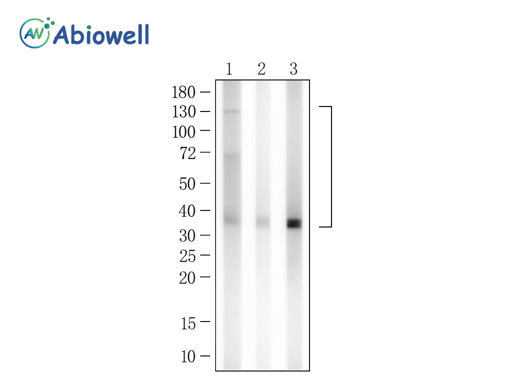
Fig : Western blot analysis of C3 on different lysates. Proteins were transferred to a NC membrane and blocked with 5% NF-Milk in TBST for 1 hour at room temperature. The primary antibody (AWA12697, 1/1000) was used in TBST at room temperature for 2 hours. Goat Anti- Rabbit IgG - HRP Secondary Antibody (AWS0002) at 1:5,000 dilution was used for 1 hour at room temperature. Positive control: Lane 1: HepG2 cell Lane 2: Mouse liver Lane 3: Rat liver Predicted molecular weight:187 kDa Observed molecular weight:130/60/37 kDa Exposure time:45s |
-
-
- 20μL
- ¥620
- 1-3个工作日
-
- 50μL
- ¥1250
- 1-3个工作日
-
- 100μL
- ¥2200
- 1-3个工作日
-
相关产品
-
Cdk6 Recombinant Rabbit Monoclonal Antibody
GAPDH Rabbit Polyclonal Antibody
GFAP Recombinant Mouse Monoclonal Antibody
Ki67 Rabbit Monoclonal Antibody
HMGB1 Recombinant Rabbit Monoclonal Antibody
SQSTM1/p62 Mouse Monoclonal Antibody
Bcl-2 Recombinant Rabbit Monoclonal Antibody
SOD2 Rabbit Polyclonal Antibody


