
PD-L1 Recombinant Rabbit Monoclonal Antibody
-
-
- 20μL
- ¥620
- 1-3个工作日
-
- 50μL
- ¥1250
- 1-3个工作日
-
- 100μL
- ¥2200
- 1-3个工作日
Product Details | Host Species: Rabbit | Reactivity: Human,Rat | Molecular Wt: 33 kDa | |
Clonality: Monoclonal | Isotype: IgG | Concentration: 1mg/ml | ||
Other Names: B7 H; B7 H1; B7 homolog 1; B7H1; CD274; CD274 molecule; PD L1; PDCD1 ligand 1; PDCD1L1; PDCD1LG1; PDL1; PD-L1; PD-L1/CD274; Programmed death ligand 1 | ||||
Formulation: Liquid in PBS containing 50% glycerol, 0.5% BSA and 0.02% sodium azide. | ||||
Purification: Affinity-chromatography | ||||
Storage: Store at -20°C. Stable for one year after shipment. Aliquoting is unnecessary for -20°C storage. | ||||
Applications | WB 1:1000-1:2000 | |||
Immunogen Information | Gene Name: CD274 | Protein Name: Programmed cell death 1 ligand 1 | ||
Gene ID: 29126 (Human) | SwissPro: Q9NZQ7 (Human) | |||
Subcellular Location: Cell membrane. Early endosome membrane. Recycling endosome membrane. Nucleus. Endomembrane system. Secreted. | ||||
Immunogen: Synthetic peptide within human PD-L1. AA range: 260-290. | ||||
Specificity: PD-L1 Monoclonal Antibody detects endogenous levels of PD-L1 protein. | ||||
| Product images | |
|
|
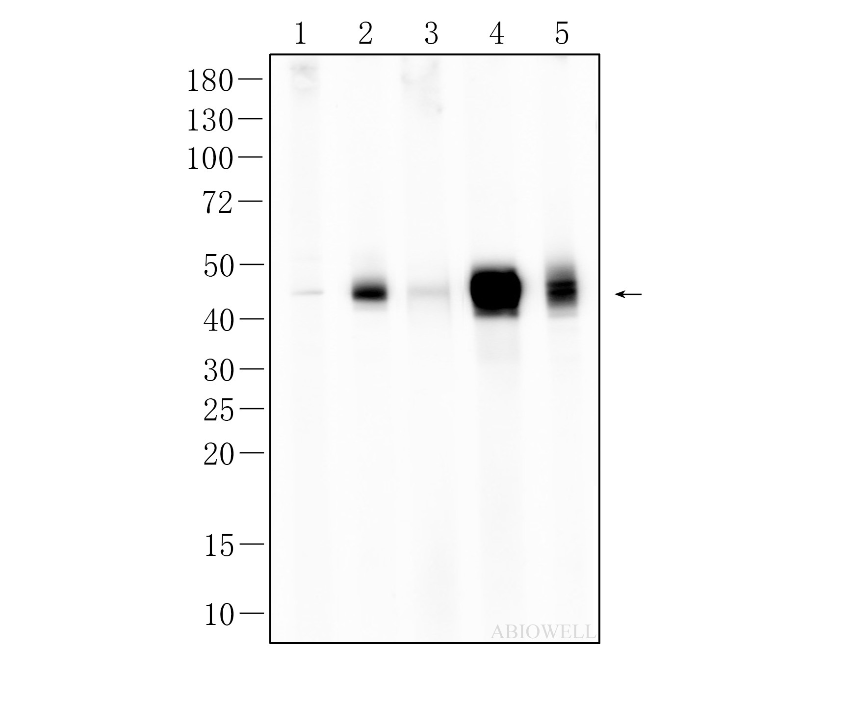
Fig : Western blot analysis of PD-L1 on different lysates. Proteins were transferred to a NC membrane and blocked with 5% NF-Milk in TBST for 1 hour at room temperature. The primary antibody (AWA12695, 1/1000) was used in TBST at room temperature for 2 hours. Goat Anti-Rabbit IgG - HRP Secondary Antibody (AWS0002) at 1:5,000 dilution was used for 1 hour at room temperature. Positive control: Lane 1: PC3 cell Lane 2: A375 cell Lane 3: A549 cell Lane 4: U87-MG cell Lane 5: Hela cell Exposure time: 7 min Predicted band size: 33 kDa Observed band size: 45-50 kDa |
-
-
- 20μL
- ¥620
- 1-3个工作日
-
- 50μL
- ¥1250
- 1-3个工作日
-
- 100μL
- ¥2200
- 1-3个工作日
-
相关产品
-
Cdk6 Recombinant Rabbit Monoclonal Antibody
GAPDH Rabbit Polyclonal Antibody
GFAP Recombinant Mouse Monoclonal Antibody
Ki67 Rabbit Monoclonal Antibody
HMGB1 Recombinant Rabbit Monoclonal Antibody
SQSTM1/p62 Mouse Monoclonal Antibody
Bcl-2 Recombinant Rabbit Monoclonal Antibody
SOD2 Rabbit Polyclonal Antibody


