
Ferritin Recombinant Rabbit Monoclonal Antibody

-
-
- 20μL
- ¥620
- 1-3个工作日
-
- 50μL
- ¥1250
- 1-3个工作日
-
- 100μL
- ¥2200
- 1-3个工作日
Product Details | Host Species: Rabbit | Reactivity: Human,Mouse,Rat,Zebrafish | Molecular Wt: 21 kDa | |
Clonality: Monoclonal | Isotype: IgG | Concentration: 1mg/ml | ||
Other Names: Cell proliferation-inducing gene 15 protein; Ferritin H subunit; Ferritin heavy chain; Ferritin heavy polypeptide 1; Ferritin L subunit; Ferritin, heavy polypeptide; FTH; FTH1; FTL | ||||
Formulation: Liquid in PBS containing 50% glycerol, 0.5% BSA and 0.02% sodium azide. | ||||
Purification: Affinity-chromatography | ||||
Storage: Store at -20°C. Stable for one year after shipment. Aliquoting is unnecessary for -20°C storage. | ||||
Applications | WB 1:1000-1:5000 | |||
Immunogen Information | Gene Name: FTH1 | Protein Name: Ferritin heavy chain | ||
Gene ID: 2495 (Human) | SwissPro: P02794 (Human) | |||
Subcellular Location: Cytoplasm. Lysosome. Cytoplasmic vesicle, autophagosome. | ||||
Immunogen: Synthetic peptide within human FTH1. AA range:range:58-99. | ||||
Specificity: 0 | ||||
| Product images | |
|
|
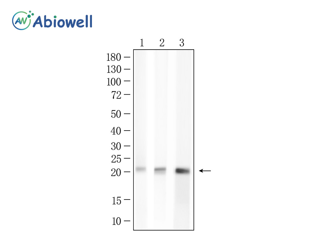
Fig : Western blot analysis of Ferritin on different lysates. Proteins were transferred to a NC membrane and blocked with 5% NF-Milk in TBST for 1 hour at room temperature. The primary antibody (AWA12692, 1/2000) was used in TBST at room temperature for 2 hours. Goat Anti-Ribbit IgG - HRP Secondary Antibody (AWS0002) at 1:5,000 dilution was used for 1 hour at room temperature. Positive control: Lane 1: Hela cell Lane 2: A549 cell Lane 3: HepA1-6 cell Exposure time: 15seconds Predicted molecular weight: 21 kDa Observed molecular weight: 21 kDa |
|
|
|
|
|
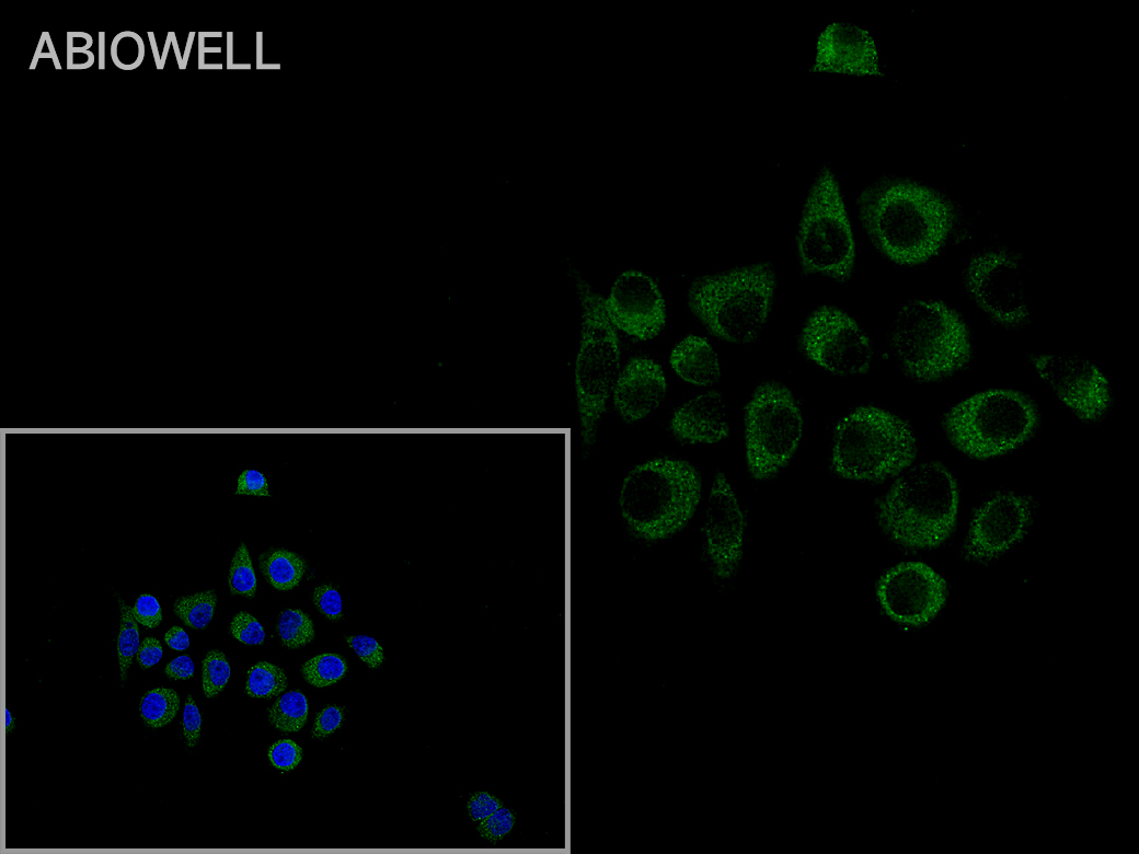
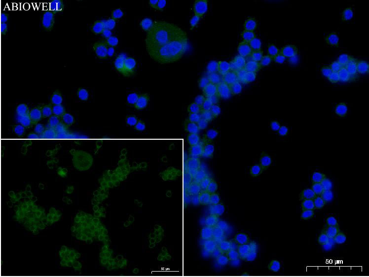
Fig: Immunocytochemistry analysis of Hela cells labeling Ferritin with Rabbit anti-Ferritin antibody (AWA12692) at 1/50 dilution(Green). Cells were fixed in 4% paraformaldehyde for 10 minutes at 37 ℃, permeabilized with 0.03% Triton X-100 in PBS for 30 minutes, and then blocked with 5% BSA for 60 minutes at 37 ℃. Cells were then incubated with Rabbit anti-Ferritin antibody (AWA12692) at 1/50 dilution in 2% negative goat serum overnight at 4 ℃. Goat Anti-Rabbit IgG H&L (iFluor™ 488, AWS0005) was used as the secondary antibody at 1/200 dilution for 60 minutes at 37 ℃. Nuclear DNA was labelled in blue with DAPI(AWC0291). |
|
|
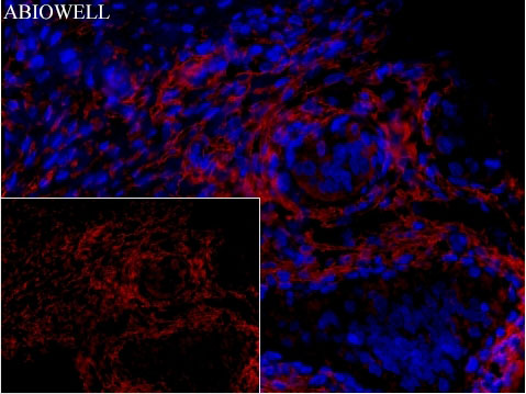
Fig: Fluorescence immunohistochemical analysis of Rat-ovary tissue (Formalin/PFA-fixed paraffin-embedded sections) with Rabbit anti-Ferritin antibody (AWA12692) at 1/100 dilution. The immunostaining was performed with the TSA Immuno-staining Kit (ABIOWELL, AWI0689). The section was pre-treated using heat mediated antigen retrieval with EDTA buffer (pH 9.0) for 20 minutes. The tissues were blocked in 1% BSA for 20 minutes at room temperature, washed with ddH2O and PBS, and then probed with the primary antibody (AWA12692) at 1/100 dilution for 1 hour at room temperature. The detection was performed using an HRP conjugated compact polymer system followed by a separate fluorescent tyramide signal amplification system (red). DAPI (blue, AWC0291) was used as a nuclear counter stain. Image acquisition was performed with Slide Scanner. |
|
|
Fig: Immunocytochemistry analysis of Hela cells labeling Ferritin with Rabbit anti-Ferritin antibody (AWA12692)at 1/50 dilution(Green). Cells were fixed in 4% paraformaldehyde for 10 minutes at 37 ℃, permeabilized with 0.03% Triton X-100 in PBS for 30 minutes, and then blocked with 5% BSA for 60 minutes at 37 ℃. Cells were then incubated with Rabbit anti-Ferritin antibody (AWA12692)at 1/50 dilution in 2% negative goat serum overnight at 4 ℃. Goat Anti-Rabbit IgG H&L (iFluor™ 488, AWS0005c) was used as the secondary antibody at 1/200 dilution for 60 minutes at 37 ℃. Nuclear DNA was labelled in blue with DAPI(AWC0291). |
|
|
Fig: Immunocytochemistry analysis of Raw264.7 cells labeling Ferritin with Rabbit anti-Ferritin antibody (AWA12692)at 1/50 dilution(Green). Cells were fixed in 4% paraformaldehyde for 10 minutes at 37 ℃, permeabilized with 0.03% Triton X-100 in PBS for 30 minutes, and then blocked with 5% BSA for 60 minutes at 37 ℃. Cells were then incubated with Rabbit anti-Ferritin antibody (AWA12692 )at 1/50 dilution in 2% negative goat serum overnight at 4 ℃. Goat Anti-Rabbit IgG H&L (iFluor™ 488, AWS0005c) was used as the secondary antibody at 1/200 dilution for 60 minutes at 37 ℃. Nuclear DNA was labelled in blue with DAPI(AWC0291). |
|
|
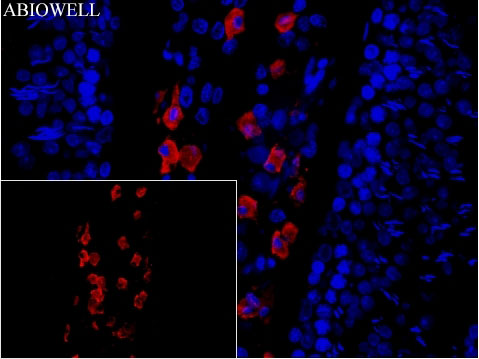
Fig: Fluorescence immunohistochemical analysis of Mouse-testicle tissue (Formalin/PFA-fixed paraffin-embedded sections) with Rabbit anti-Ferritin antibody (AWA12692) at 1/100 dilution. The immunostaining was performed with the TSA Immuno-staining Kit (ABIOWELL, AWI0689). The section was pre-treated using heat mediated antigen retrieval with EDTA buffer (pH 9.0) for 20 minutes. The tissues were blocked in 1% BSA for 20 minutes at room temperature, washed with ddH2O and PBS, and then probed with the primary antibody (AWA12692) at 1/100 dilution for 1 hour at room temperature. The detection was performed using an HRP conjugated compact polymer system followed by a separate fluorescent tyramide signal amplification system (red). DAPI (blue, AWC0291) was used as a nuclear counter stain. Image acquisition was performed with Slide Scanner. |
|
|
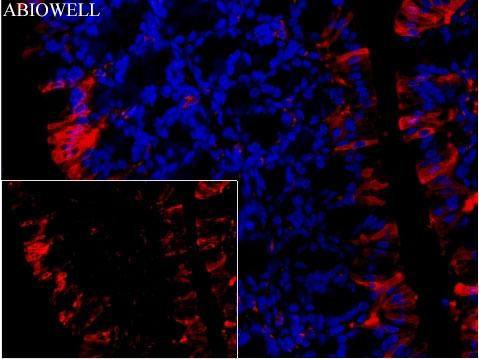
Fig: Fluorescence immunohistochemical analysis of Mouse-colon tissue (Formalin/PFA-fixed paraffin-embedded sections) with Rabbit anti-Ferrintin antibody (AWA12692) at 1/100 dilution. The immunostaining was performed with the TSA Immuno-staining Kit (ABIOWELL, AWI0689). The section was pre-treated using heat mediated antigen retrieval with EDTA buffer (pH 9.0) for 20 minutes. The tissues were blocked in 1% BSA for 20 minutes at room temperature, washed with ddH2O and PBS, and then probed with the primary antibody (AWA12692) at 1/100 dilution for 1 hour at room temperature. The detection was performed using an HRP conjugated compact polymer system followed by a separate fluorescent tyramide signal amplification system (red). DAPI (blue, AWC0291) was used as a nuclear counter stain. Image acquisition was performed with Slide Scanner. |
|
|
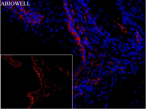
Fig: Fluorescence immunohistochemical analysis of Mouse-stomach tissue (Formalin/PFA-fixed paraffin-embedded sections) with Rabbit anti-Ferritin antibody (AWA12692) at 1/100 dilution. The immunostaining was performed with the TSA Immuno-staining Kit (ABIOWELL, AWI0689). The section was pre-treated using heat mediated antigen retrieval with EDTA buffer (pH 9.0) for 20 minutes. The tissues were blocked in 1% BSA for 20 minutes at room temperature, washed with ddH2O and PBS, and then probed with the primary antibody (AWA12692) at 1/100 dilution for 1 hour at room temperature. The detection was performed using an HRP conjugated compact polymer system followed by a separate fluorescent tyramide signal amplification system (red). DAPI (blue, AWC0291) was used as a nuclear counter stain. Image acquisition was performed with Slide Scanner. |
|
|
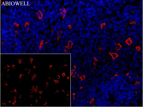
Fig: Fluorescence immunohistochemical analysis of Rat-spleen tissue (Formalin/PFA-fixed paraffin-embedded sections). with Rabbit anti-Ferritin antibody (AWA12692) at 1/100 dilution. The immunostaining was performed with the TSA Immuno-staining Kit (ABIOWELL, AWI0689). The section was pre-treated using heat mediated antigen retrieval with EDTA buffer (pH 9.0) for 20 minutes. The tissues were blocked in 1% BSA for 20 minutes at room temperature, washed with ddH2O and PBS, and then probed with the primary antibody (AWA12692) at 1/100 dilution for 1 hour at room temperature. The detection was performed using an HRP conjugated compact polymer system followed by a separate fluorescent tyramide signal amplification system (red). DAPI (blue, AWC0291) was used as a nuclear counter stain. Image acquisition was performed with Slide Scanner. |
-
-
- 20μL
- ¥620
- 1-3个工作日
-
- 50μL
- ¥1250
- 1-3个工作日
-
- 100μL
- ¥2200
- 1-3个工作日
-
相关产品
-
Cdk6 Recombinant Rabbit Monoclonal Antibody
GAPDH Rabbit Polyclonal Antibody
GFAP Recombinant Mouse Monoclonal Antibody
Ki67 Rabbit Monoclonal Antibody
HMGB1 Recombinant Rabbit Monoclonal Antibody
SQSTM1/p62 Mouse Monoclonal Antibody
Bcl-2 Recombinant Rabbit Monoclonal Antibody
SOD2 Rabbit Polyclonal Antibody


