
STAT3 Recombinant Rabbit Monoclonal Antibody
-
-
- 20μL
- ¥620
- 有库存
-
- 50μL
- ¥1250
- 有库存
-
- 100μL
- ¥2200
- 有库存
Product Details
| Host Species: Rabbit | Reactivity: Human,Mouse,Rat | Molecular Wt: 88 kDa | |
Clonality: Monoclonal | Isotype: IgG | Concentration: 1 mg/ml | ||
Other Names: STAT3; APRF; Signal transducer and activator of transcription 3; Acute-phase response factor
| ||||
Formulation: Liquid in PBS containing 50% glycerol, 0.5% BSA and 0.02% sodium azide. | ||||
Purification: Affinity-chromatography | ||||
Storage: -20°C,1 year | ||||
Applications
| WB 1:500-1:2000 IHC-P 1:50-1:1000 IF 1:100-1:500
| |||
Immunogen Information | Gene Name: STAT3 | Protein Name: Signal transducer and activator of transcription 3 | ||
Gene ID: 6774 (Human) 20848 (Mouse) 25125 (Rat) | SwissPro: P40763 (Human) P42227 (Mouse) P52631 (Rat)
| |||
Immunogen: Synthetic peptide within C-terminal human STAT3. | ||||
Specificity: STAT3 Monoclonal Antibody detects endogenous levels of STAT3 protein. | ||||
| Product images | |
|
|
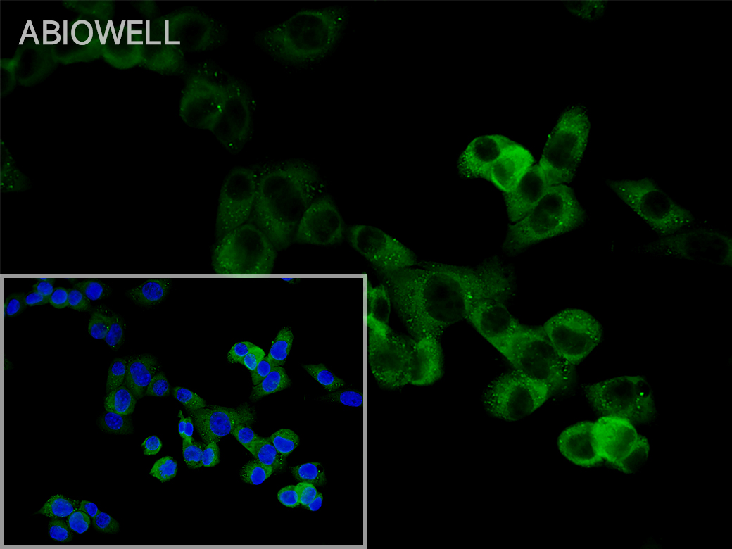
Fig: Immunocytochemistry analysis of NIH3T3 cells labeling STAT3 with Rabbit anti-STAT3 antibody (AWA12672) at 1/50 dilution(green). Cells were fixed in 4% paraformaldehyde for 10 minutes at 37 ℃, permeabilized with 0.03% Triton X-100 in PBS for 30 minutes, and then blocked with 5% BSA for 60 minutes at 37 ℃. Cells were then incubated with Rabbit anti-STAT3 antibody (AWA12672) at 1/50 dilution in 2% negative goat serum overnight at 4 ℃. Goat Anti-Rabbit IgG H&L (iFluor™ 488, AWS0005c) was used as the secondary antibody at 1/200 dilution for 60 minutes at 37 ℃. Nuclear DNA was labelled in blue with DAPI(AWC0291). |
|
|
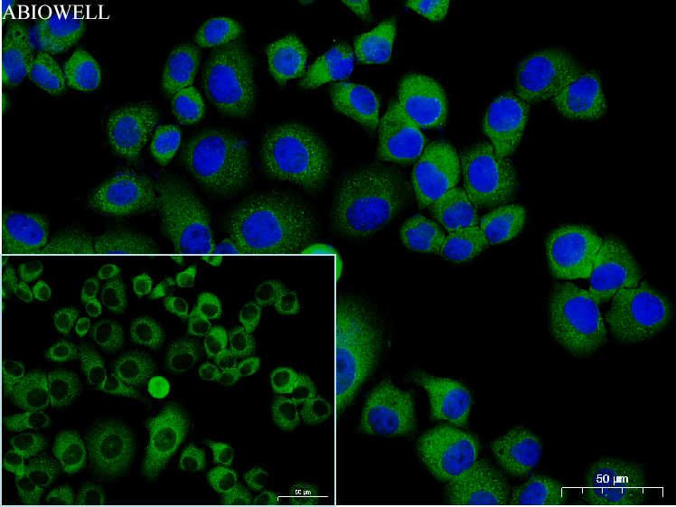
Fig: Immunocytochemistry analysis of Hela cells labeling STAT3 with Rabbit anti-STAT3 antibody (AWA12672)at 1/100 dilution(Green). Cells were fixed in 4% paraformaldehyde for 10 minutes at 37 ℃, permeabilized with 0.03% Triton X-100 in PBS for 30 minutes, and then blocked with 5% BSA for 60 minutes at 37 ℃. Cells were then incubated with Rabbit anti-STAT3 antibody (AWA12672)at 1/100 dilution in 2% negative goat serum overnight at 4 ℃. Goat Anti-Rabbit IgG H&L (iFluor™ 488, AWS0005c) was used as the secondary antibody at 1/200 dilution for 60 minutes at 37 ℃. Nuclear DNA was labelled in blue with DAPI(AWC0291). |
|
|
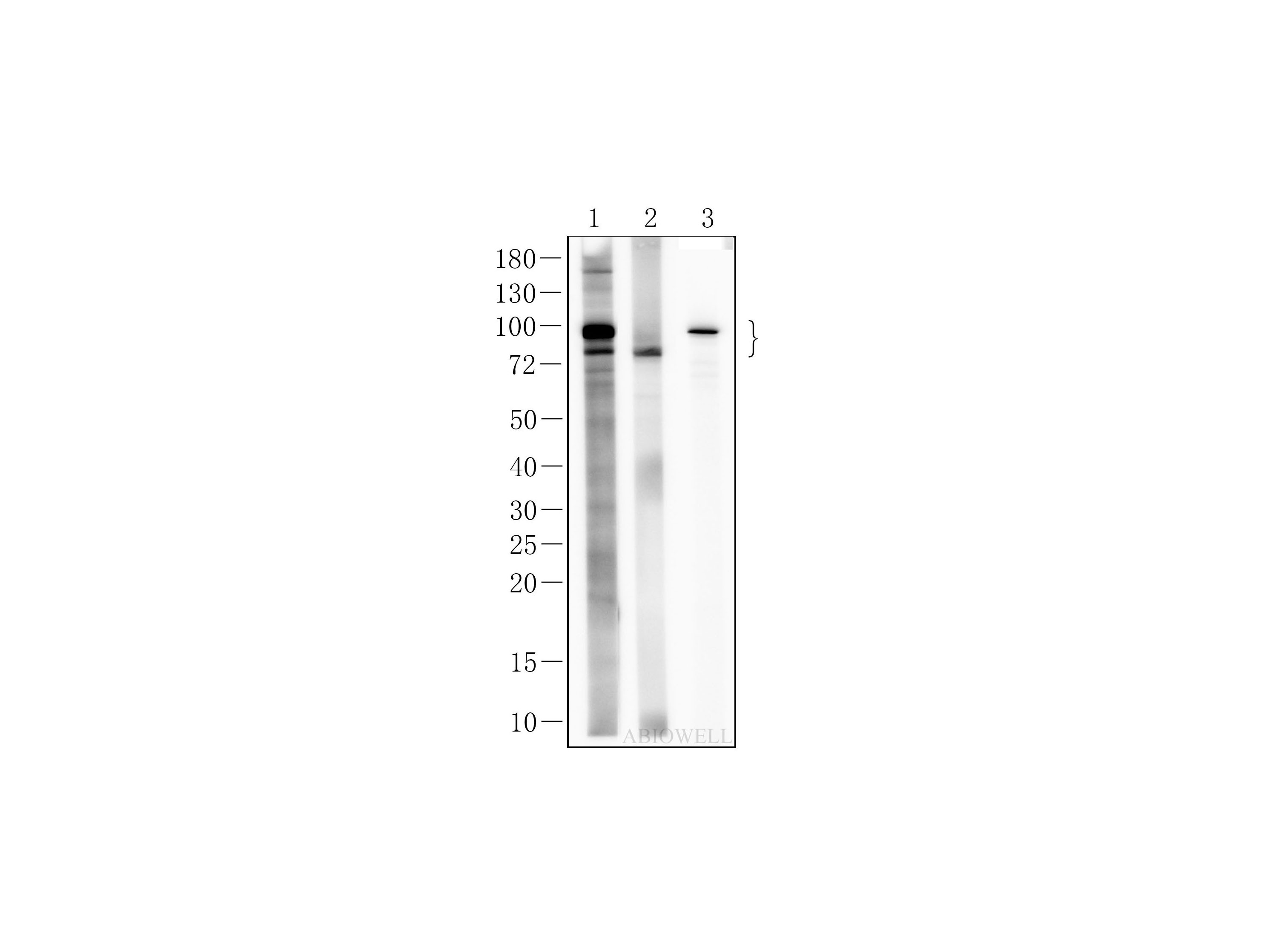
Fig : Western blot analysis of STAT3 on different lysates. Proteins were transferred to a NC membrane and blocked with 5% NF-Milk in TBST for 1 hour at room temperature. The primary antibody (AWA12672, 1/1000) was used in TBST at room temperature for 2 hours. Goat Anti-rabbit IgG - HRP Secondary Antibody (AWS0001) at 1:5,000 dilution was used for 1 hour at room temperature. Positive control: Lane 1:Hela cell lysate Lane 2: Rat brain tissue lysate Lane 3: HUVEC cell lysate |
|
|
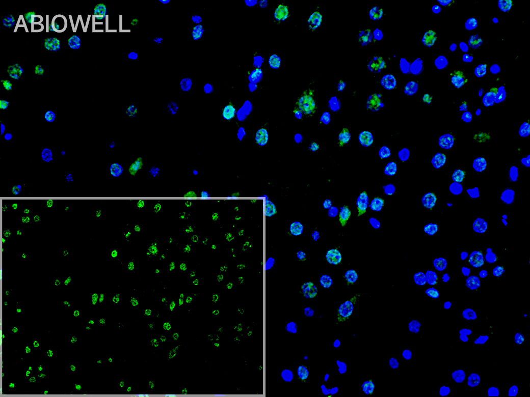
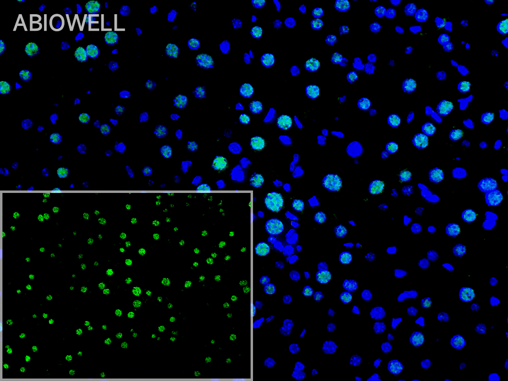
Fig: Fluorescence immunohistochemical analysis of Rat-Brain tissue (Formalin/PFA-fixed paraffin-embedded sections). with Rabbit anti-STAT3 antibody (AWA12672) at 1/200 dilution. The immunostaining was performed with the TSA Immuno-staining Kit (ABIOWELL, AWI0688). The section was pre-treated using heat mediated antigen retrieval with EDTA buffer (pH 9.0) for 20 minutes. The tissues were blocked in 5% BSA for 60 minutes at 37℃, washed with ddH2O and PBS, and then probed with the primary antibody (AWA12672) at 1/200 dilution for 1 hour at room temperature. The detection was performed using an HRP conjugated compact polymer system followed by a separate fluorescent tyramide signal amplification system (green). DAPI (blue, AWC0291) was used as a nuclear counter stain. Image acquisition was performed with Slide Scanner. |
|
|
Fig: Fluorescence immunohistochemical analysis of Mouse-liver tissue (Formalin/PFA-fixed paraffin-embedded sections). with Rabbit anti-STAT3 antibody (AWA12672) at 1/200 dilution. The immunostaining was performed with the TSA Immuno-staining Kit (ABIOWELL, AWI0688). The section was pre-treated using heat mediated antigen retrieval with EDTA buffer (pH 9.0) for 20 minutes. The tissues were blocked in 5% BSA for 60 minutes at 37℃, washed with ddH2O and PBS, and then probed with the primary antibody (AWA12672) at 1/200 dilution for 1 hour at room temperature. The detection was performed using an HRP conjugated compact polymer system followed by a separate fluorescent tyramide signal amplification system (green). DAPI (blue, AWC0291) was used as a nuclear counter stain. Image acquisition was performed with Slide Scanner. |
-
-
- 20μL
- ¥620
- 1-3个工作日
-
- 50μL
- ¥1250
- 1-3个工作日
-
- 100μL
- ¥2200
- 1-3个工作日
-
相关产品
-
Cdk6 Recombinant Rabbit Monoclonal Antibody
GAPDH Rabbit Polyclonal Antibody
GFAP Recombinant Mouse Monoclonal Antibody
Ki67 Rabbit Monoclonal Antibody
HMGB1 Recombinant Rabbit Monoclonal Antibody
SQSTM1/p62 Mouse Monoclonal Antibody
Bcl-2 Recombinant Rabbit Monoclonal Antibody
SOD2 Rabbit Polyclonal Antibody


