
CD31 Recombinant Rabbit Monoclonal Antibody
-
-
- 20μL
- ¥620
- 1-3个工作日
-
- 50μL
- ¥1250
- 1-3个工作日
-
- 100μL
- ¥2200
- 1-3个工作日
Product Details | Host Species: Rabbit | Reactivity: Human,Mouse,Rat | Molecular Wt: 83 kDa | |
Clonality: Monoclonal | Isotype: IgG | Concentration: 1mg/ml | ||
Other Names: PECAM1; Platelet endothelial cell adhesion molecule; PECAM-1; EndoCAM; GPIIA'; PECA1; CD antigen CD31 | ||||
Formulation: Liquid in PBS containing 50% glycerol, 0.5% BSA and 0.02% sodium azide. | ||||
Purification: Affinity-chromatography | ||||
Storage: Store at -20°C. Stable for one year after shipment. Aliquoting is unnecessary for -20°C storage. | ||||
Applications | WB 1:1000-1:2000 | |||
Immunogen Information | Gene Name: PECAM1 | Protein Name: Platelet endothelial cell adhesion molecule | ||
Gene ID: 5175 (Human) | SwissPro: P16284 (Human) | |||
Subcellular Location: Cell membrane. Membrane raft. Cell junction. | ||||
Immunogen: Synthetic peptide within human CD31. AA range: 689-738. | ||||
Specificity: CD31 Monoclonal Antibody detects endogenous levels of CD31 protein. | ||||
| Product images | |
|
|
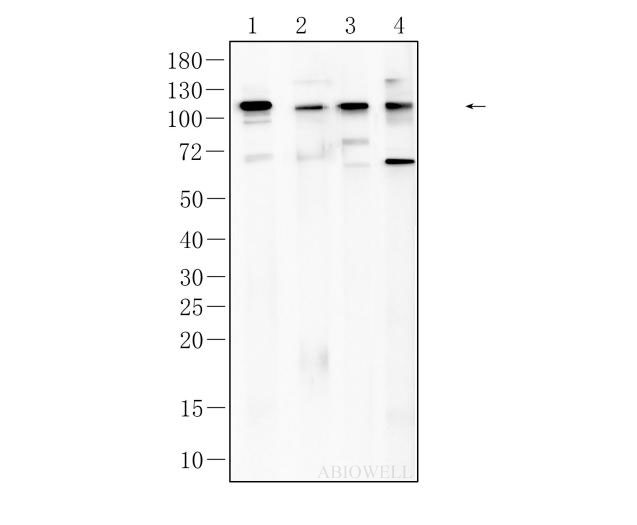
Fig : Western blot analysis of CD31 on different lysates. Proteins were transferred to a NC membrane and blocked with 5% NF-Milk in TBST for 1 hour at room temperature. The primary antibody (AWA11937, 1/1000) was used in TBST at room temperature for 2 hours. Goat Anti-Rabbit IgG - HRP Secondary Antibody (AWS0002) at 1:5,000 dilution was used for 1 hour at room temperature. Positive control: Lane 1:Jurkat cell Lane 2: HEK-293 cell Lane 3:NIH3T3 cell Lane 4: RBL-2H3 cell Predicted band size: 83 kDa Observed band size: 110 kDa |
|
|
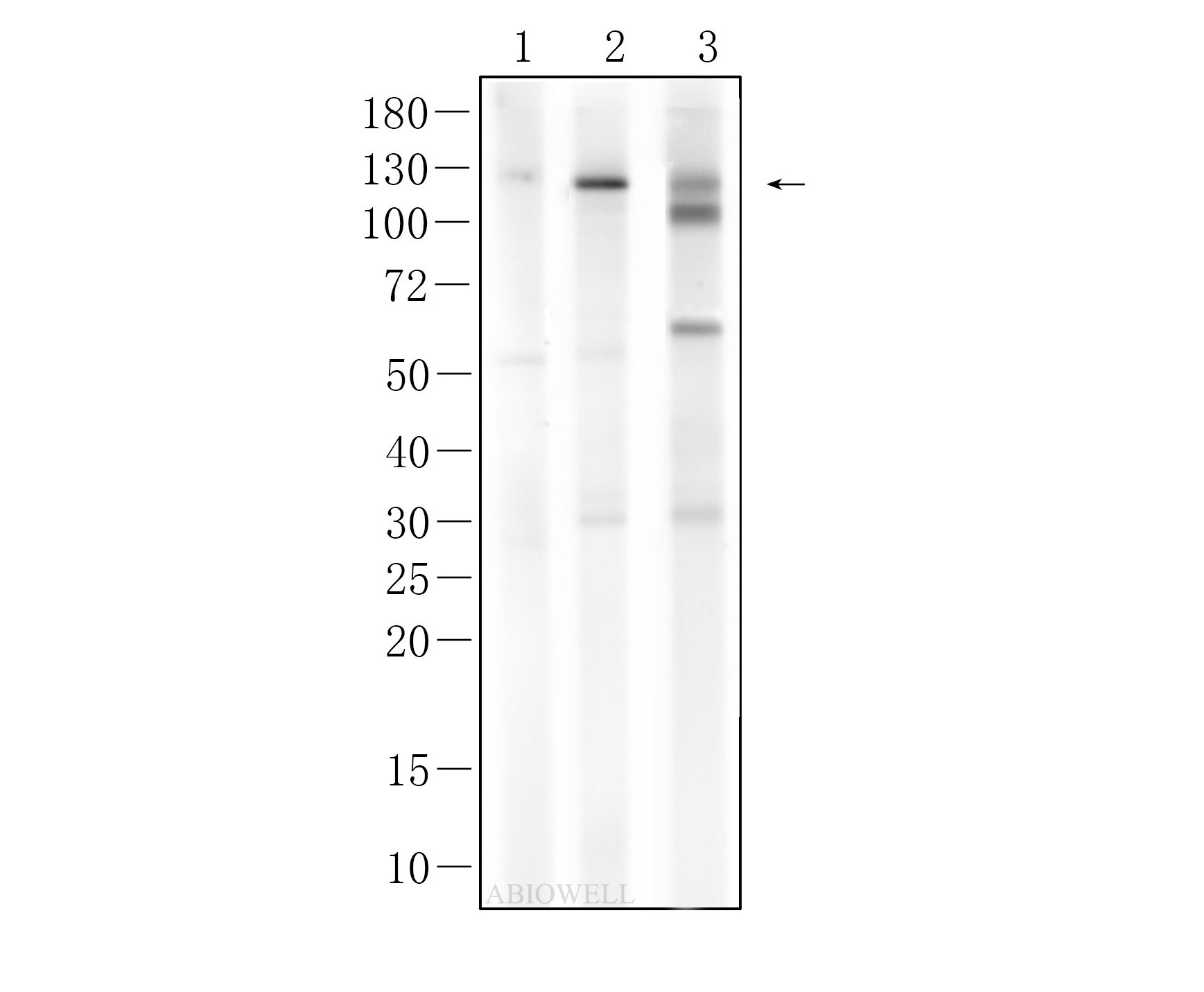
Fig : Western blot analysis of CD31 on different lysates. Proteins were transferred to a NC membrane and blocked with 5% NF-Milk in TBST for 1 hour at room temperature. The primary antibody (AWA11937, 1/1000) was used in TBST at room temperature for 2 hours. Goat Anti-Rabbit IgG - HRP Secondary Antibody (AWS0002) at 1:5,000 dilution was used for 1 hour at room temperature. Positive control: Lane 1: RBL-2H3 cell Lane 2: Jurkat cell Lane 3: NIH3T3 cell Exposure time: 90 seconds Predicted molecular weight:83 kDa Observed molecular weight:110 kDa |
|
|
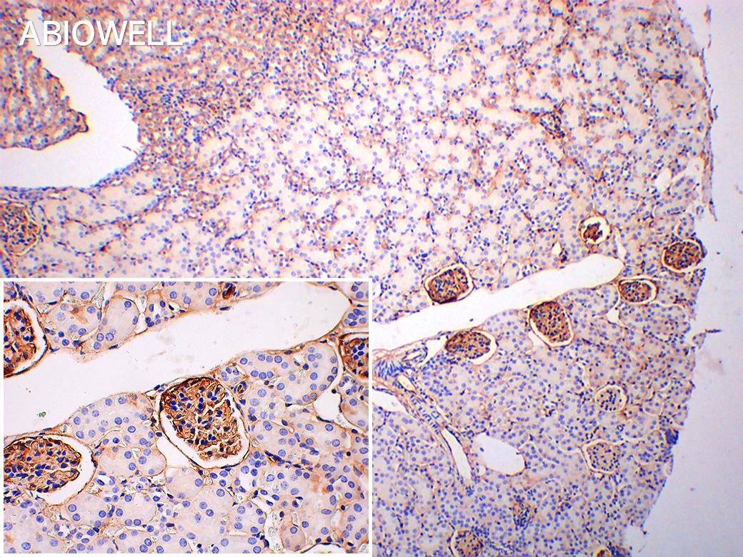
Fig : Immunohistochemical analysis of paraffin-embedded Mouse-kidney tissue with Rabbit anti-CD31 antibody (AWA11937) at 1/200 dilution. The section was pre-treated using heat mediated antigen retrieval with Sodium citrate buffer (pH 6.0) for 20 minutes. The tissues were blocked in 3% H2O2 for 15 minutes at room temperature, washed with ddH2O and PBS, and then probed with the primary antibody (AWA11937) at 1/200 dilution for 2 hour at 37℃or overnignt at 4℃. The detection was performed using an HRP conjugated compact polymer system(ABIOWELL, AWI0629). DAB was used as the chromogen. Tissues were counterstained with hematoxylin and mounted with DPX. |
-
-
- 20μL
- ¥620
- 1-3个工作日
-
- 50μL
- ¥1250
- 1-3个工作日
-
- 100μL
- ¥2200
- 1-3个工作日
-
相关产品
-
Cdk6 Recombinant Rabbit Monoclonal Antibody
GAPDH Rabbit Polyclonal Antibody
GFAP Recombinant Mouse Monoclonal Antibody
Ki67 Rabbit Monoclonal Antibody
HMGB1 Recombinant Rabbit Monoclonal Antibody
SQSTM1/p62 Mouse Monoclonal Antibody
Bcl-2 Recombinant Rabbit Monoclonal Antibody
SOD2 Rabbit Polyclonal Antibody


