
P53 Mouse Monoclonal Antibody
-
-
- 20μL
- ¥620
- 1-3个工作日
-
- 50μL
- ¥1250
- 1-3个工作日
-
- 100μL
- ¥2200
- 1-3个工作日
Product Details
| Host Species: Mouse | Reactivity: Human,Rat | Molecular Wt: Predicted MW: 44 kDa Observed MW: 53 kDa
| |
Clonality: Monoclonal | Isotype: IgG2a | |||
Other Names: TP53; P53; Cellular tumor antigen p53; Antigen NY-CO-13; Phosphoprotein p53; Tumor suppressor p53; BCC7; LFS1; BMFS5; TRP53
| ||||
Formulation: Liquid in PBS containing 50% glycerol, 0.5% BSA and 0.02% sodium azide. | ||||
Purification: Affinity-chromatography | ||||
Storage: -20°C,1 year | ||||
Applications
| WB 1:500-1:2000 | |||
Immunogen Information | Gene Name: TP53 | Protein Name: Cellular tumor antigen p53 | ||
Gene ID: 7157 (Human) 24842 (Rat) | SwissPro: P04637 (Human) P10361 (Rat)
| |||
Subcellular Location: Cytoplasm. Nucleus. Nucleus, PML body. Endoplasmic reticulum. Mitochondrion matrix. Cytoplasm, cytoskeleton, microtubule organizing center, centrosome.
| ||||
Immunogen: Synthesized peptide derived from human P53. AA range: 250-393. | ||||
Specificity: P53 Monoclonal Antibody detects endogenous levels of P53 protein. | ||||
| Product images | |
|
|
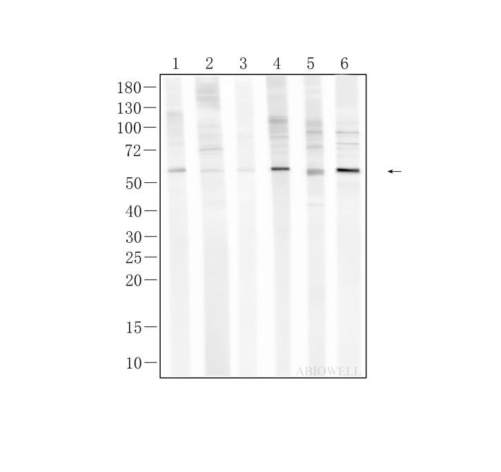
Fig : Western blot analysis of P53 on different lysates. Proteins were transferred to a NC membrane and blocked with 5% NF-Milk in TBST for 1 hour at room temperature. The primary antibody (AWA00214, 1/1000) was used in TBST at room temperature for 2 hours. Goat Anti-Mouse IgG - HRP Secondary Antibody (AWS0002) at 1:5,000 dilution was used for 1 hour at room temperature. Positive control: Lane 1: PC-12 cell Lane 2:A2058 cell Lane 3: A375 cell Lane 4: RBL-2H3 cell Lane 5: HCT116 cell Lane 6: HEK-293 cell Calculated molecular weight: 44kDa Observed molecular weight: 53kDa |
-
-
- 20μL
- ¥620
- 1-3个工作日
-
- 50μL
- ¥1250
- 1-3个工作日
-
- 100μL
- ¥2200
- 1-3个工作日
-
相关产品
-
Cdk6 Recombinant Rabbit Monoclonal Antibody
GAPDH Rabbit Polyclonal Antibody
GFAP Recombinant Mouse Monoclonal Antibody
Ki67 Rabbit Monoclonal Antibody
HMGB1 Recombinant Rabbit Monoclonal Antibody
SQSTM1/p62 Mouse Monoclonal Antibody
Bcl-2 Recombinant Rabbit Monoclonal Antibody
SOD2 Rabbit Polyclonal Antibody


