
CD4 Mouse Monoclonal Antibody
-
-
- 20μL
- ¥620
- 1-3个工作日
-
- 50μL
- ¥1250
- 1-3个工作日
-
- 100μL
- ¥2200
- 1-3个工作日
Product Details
| Host Species: Mouse | Reactivity: Human,Mouse,Rat | Molecular Wt: 51 kDa | |
Clonality: Monoclonal | ||||
Other Names: CD4; T-cell surface glycoprotein CD4; T-cell surface antigen T4/Leu-3; CD antigen CD4
| ||||
Formulation: Liquid in PBS containing 50% glycerol, 0.5% BSA and 0.02% sodium azide. | ||||
Purification: Affinity-chromatography | ||||
Storage: -20°C,1 year | ||||
Applications
| IHC 1:200 | |||
Immunogen Information | Gene Name: CD4 | Protein Name: T-cell surface glycoprotein CD4 | ||
Gene ID: 920 (Human) 12504 (Mouse) 24932 (Rat)
| SwissPro: P01730 (Human) P06332 (Mouse) P05540 (Rat)
| |||
Subcellular Location: Cell membrane. | ||||
Immunogen: Synthetic Peptide of CD4. | ||||
Specificity: CD4 Monoclonal Antibody detects endogenous levels of CD4 protein. | ||||
| Product images | |
|
|
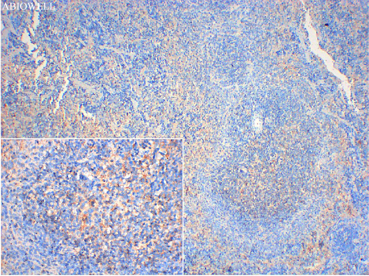
Fig : Immunohistochemical analysis of paraffin-embedded Mouse-spleen tissue with Mouse anti-CD4 antibody (AWA00070) at 1/100 dilution. The section was pre-treated using heat mediated antigen retrieval with Sodium citrate buffer (pH 6.0) for 20 minutes. The tissues were blocked in 3% H2O2 for 15 minutes at room temperature, washed with ddH2O and PBS, and then probed with the primary antibody (AWA00070) at 1/100 dilution for 1 hour at room temperature. The detection was performed using an HRP conjugated compact polymer system(ABIOWELL, AWI0629). DAB was used as the chromogen. Tissues were counterstained with hematoxylin and mounted with DPX. |
|
|
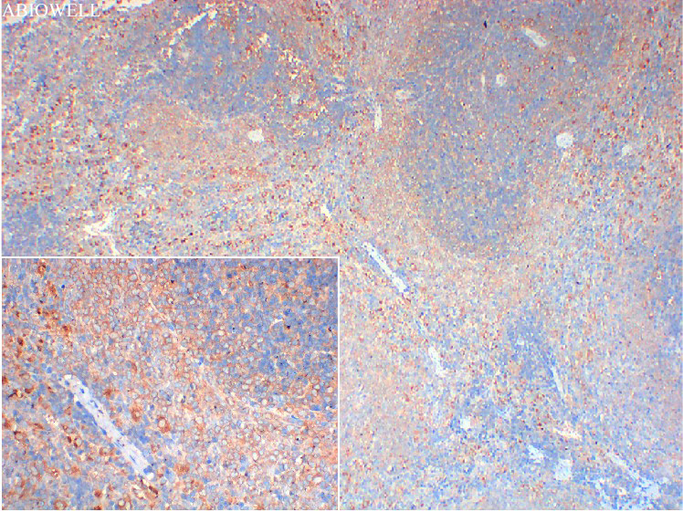
Fig : Immunohistochemical analysis of paraffin-embedded Rat-spleen tissue with Mouse anti-CD4 antibody (AWA00070) at 1/100 dilution. The section was pre-treated using heat mediated antigen retrieval with Sodium citrate buffer (pH 6.0) for 20 minutes. The tissues were blocked in 3% H2O2 for 15 minutes at room temperature, washed with ddH2O and PBS, and then probed with the primary antibody (AWA00070) at 1/100 dilution for 1 hour at room temperature. The detection was performed using an HRP conjugated compact polymer system(ABIOWELL, AWI0629). DAB was used as the chromogen. Tissues were counterstained with hematoxylin and mounted with DPX. |
-
-
- 20μL
- ¥620
- 1-3个工作日
-
- 50μL
- ¥1250
- 1-3个工作日
-
- 100μL
- ¥2200
- 1-3个工作日
-
相关产品
-
Cdk6 Recombinant Rabbit Monoclonal Antibody
GAPDH Rabbit Polyclonal Antibody
GFAP Recombinant Mouse Monoclonal Antibody
Ki67 Rabbit Monoclonal Antibody
HMGB1 Recombinant Rabbit Monoclonal Antibody
SQSTM1/p62 Mouse Monoclonal Antibody
Bcl-2 Recombinant Rabbit Monoclonal Antibody
SOD2 Rabbit Polyclonal Antibody


