
MAP2 Mouse Monoclonal Antibody
-
-
- 20μL
- ¥620
- 1-3个工作日
-
- 50μL
- ¥1250
- 1-3个工作日
-
- 100μL
- ¥2200
- 1-3个工作日
Product Details | Host Species: Mouse | Reactivity: Human,Mouse,Rat | Molecular Wt: 200 kDa | |
Clonality: Monoclonal | Isotype: IgG1 | Concentration: 1mg/ml | ||
Other Names: MAP 2; MAP-2; MAP2; Microtubule-associated protein 2; Microtubule associated protein 2; MAP2A; MAP2B; MAP2C; DKFZp686I2148; MAP dendrite specific | ||||
Formulation: Liquid in PBS containing 50% glycerol, 0.5% BSA and 0.02% sodium azide. | ||||
Purification: Affinity-chromatography | ||||
Storage: Store at -20°C. Stable for one year after shipment. Aliquoting is unnecessary for -20°C storage. | ||||
Applications | WB 1:500-2000 | |||
Immunogen Information | Gene Name: MAP2 | Protein Name: Microtubule-associated protein 2 | ||
Gene ID: 4133 (Human) | SwissPro: P11137 (Human) | |||
Subcellular Location: Cytoplasm, cytoskeleton. Cell projection, dendrite. | ||||
Immunogen: Synthetic Peptide of MAP2. | ||||
Specificity: MAP2 Monoclonal Antibody detects endogenous levels of MAP2 protein. | ||||
| Product images | |
|
|
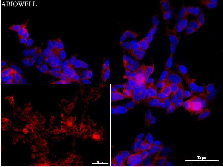
Fig: Immunocytochemistry analysis of SH-SY5Y cells labelingMAP2 with Mouse anti-MAP2 antibody (AWA00067)at 1/50 dilution(Red). Cells were fixed in 4% paraformaldehyde for 10 minutes at 37 ℃, permeabilized with 0.03% Triton X-100 in PBS for 30 minutes, and then blocked with 5% BSA for 60 minutes at 37 ℃. Cells were then incubated with Mouse anti-MAP2 antibody (AWA00067)at 1/50 dilution in 2% negative goat serum overnight at 4 ℃. Goat Anti-Mouse IgG H&L (iFluor™ 567, AWS0004c) was used as the secondary antibody at 1/200 dilution for 60 minutes at 37 ℃. Nuclear DNA was labelled in blue with DAPI(AWC0291). |
|
|
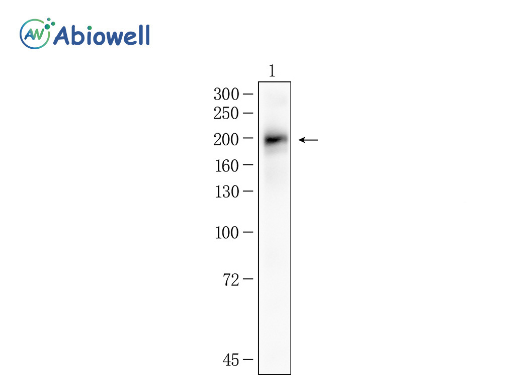
Fig : Western blot analysis of MAP2 on different lysates. Proteins were transferred to a NC membrane and blocked with 5% NF-Milk in TBST for 1 hour at room temperature. The primary antibody (AWA00067, 1/1000) was used in TBST at room temperature for 2 hours. Goat Anti-Mouse IgG - HRP Secondary Antibody (AWS0001) at 1:5,000 dilution was used for 1 hour at room temperature. Positive control: Lane 1: Hela cell Predicted molecular weight:200KD Observed molecular weight:200KD Exposure time:45 sec |
-
-
- 20μL
- ¥620
- 1-3个工作日
-
- 50μL
- ¥1250
- 1-3个工作日
-
- 100μL
- ¥2200
- 1-3个工作日
-
相关产品
-
Cdk6 Recombinant Rabbit Monoclonal Antibody
GAPDH Rabbit Polyclonal Antibody
GFAP Recombinant Mouse Monoclonal Antibody
Ki67 Rabbit Monoclonal Antibody
HMGB1 Recombinant Rabbit Monoclonal Antibody
SQSTM1/p62 Mouse Monoclonal Antibody
Bcl-2 Recombinant Rabbit Monoclonal Antibody
SOD2 Rabbit Polyclonal Antibody


