β-actin Rabbit Polyclonal Antibody
-
-
- 50μL
- ¥580
- 1-3个工作日
-
- 100μL
- ¥920
- 1-3个工作日
-
- 500μL
- ¥3800
- 1-3个工作日
Product Details
| Host Species: Rabbit | Reactivity: Human,Mouse,Rat,Chicken, Globefish,Bovine,Hamster,Pig, Ovine,Cat,Pig,Dog,Sheep
| Molecular Wt: 42 kDa | |
Clonality: Polyclonal | Isotype: IgG | Concentration: 1 mg/ml | ||
Other Names: ACTB; Actin; cytoplasmic 1; Beta-actin; Actin β; Actin Beta; β-Actin; Beta-Actin; Beta actin; PS1TP5BP1
| ||||
Formulation: Liquid in PBS containing 50% glycerol, 0.5% BSA and 0.02% sodium azide. | ||||
Purification: Affinity-chromatography | ||||
Storage: -20°C,1 year | ||||
Applications
| WB 1:1000-1:5000 IF 1:50-1:200 ELISA 1:20000
| |||
Immunogen Information | Gene Name: ACTB | Protein Name: Actin, cytoplasmic 1 | ||
Gene ID: 60 (Human) 11461 (Mouse) 81822 (Rat) | SwissPro: P60709 (Human) P60710 (Mouse) P60711 (Rat)
| |||
Subcellular Location: Cytoplasm, cytoskeleton. Nucleus. | ||||
Immunogen: Synthesized peptide derived from the N-terminal region of human Actin β. AA range: 1- 80.
| ||||
Specificity: β-actin Polyclonal Antibody detects endogenous levels of β-actin protein. | ||||
| Product images | |
|
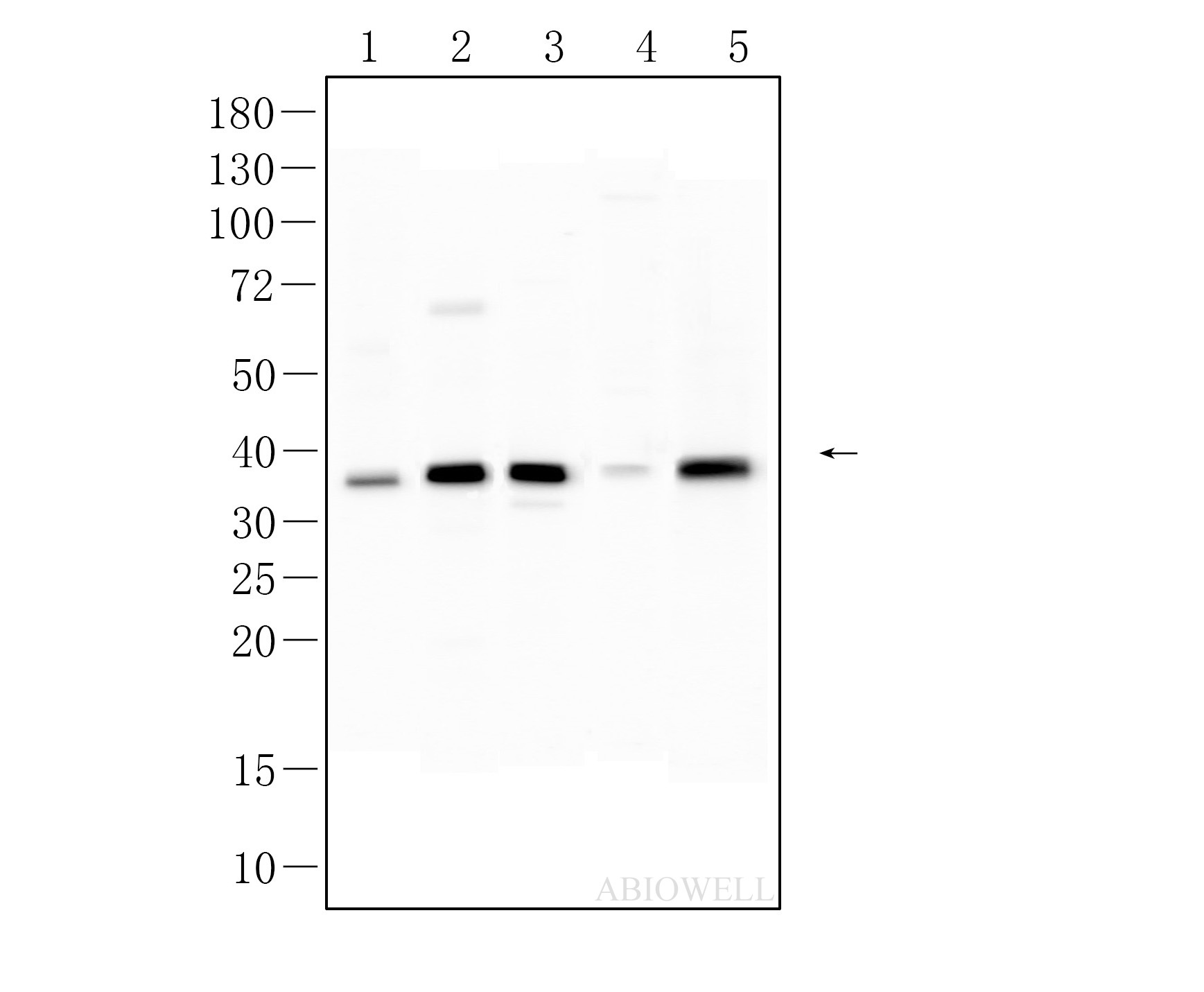
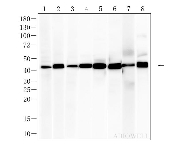
Fig : Western blot analysis of Beta actin on different lysates. Proteins were transferred to a NC membrane and blocked with 5% NF-Milk in TBST for 1 hour at room temperature. The primary antibody (AWA80002, 1/5000) was used in TBST at room temperature for 2 hours. Goat Anti-Rabbit IgG - HRP Secondary Antibody (AWS0002) at 1:5,000 dilution was used for 1 hour at room temperature. Positive control: Lane 1: HEPG2 cell Lane 2: Jurkat cell Lane 3: RAW264.7 cell Lane 4: HEK-293 cell Lane 5: A549 cell Lane 6: HELA cell Lane 7: NIH3T3 cell Lane 8: HEPA1-6 cell |
|
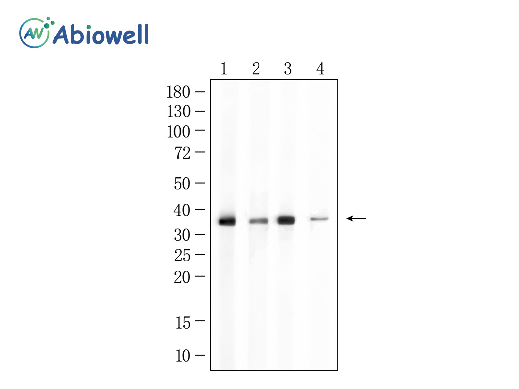
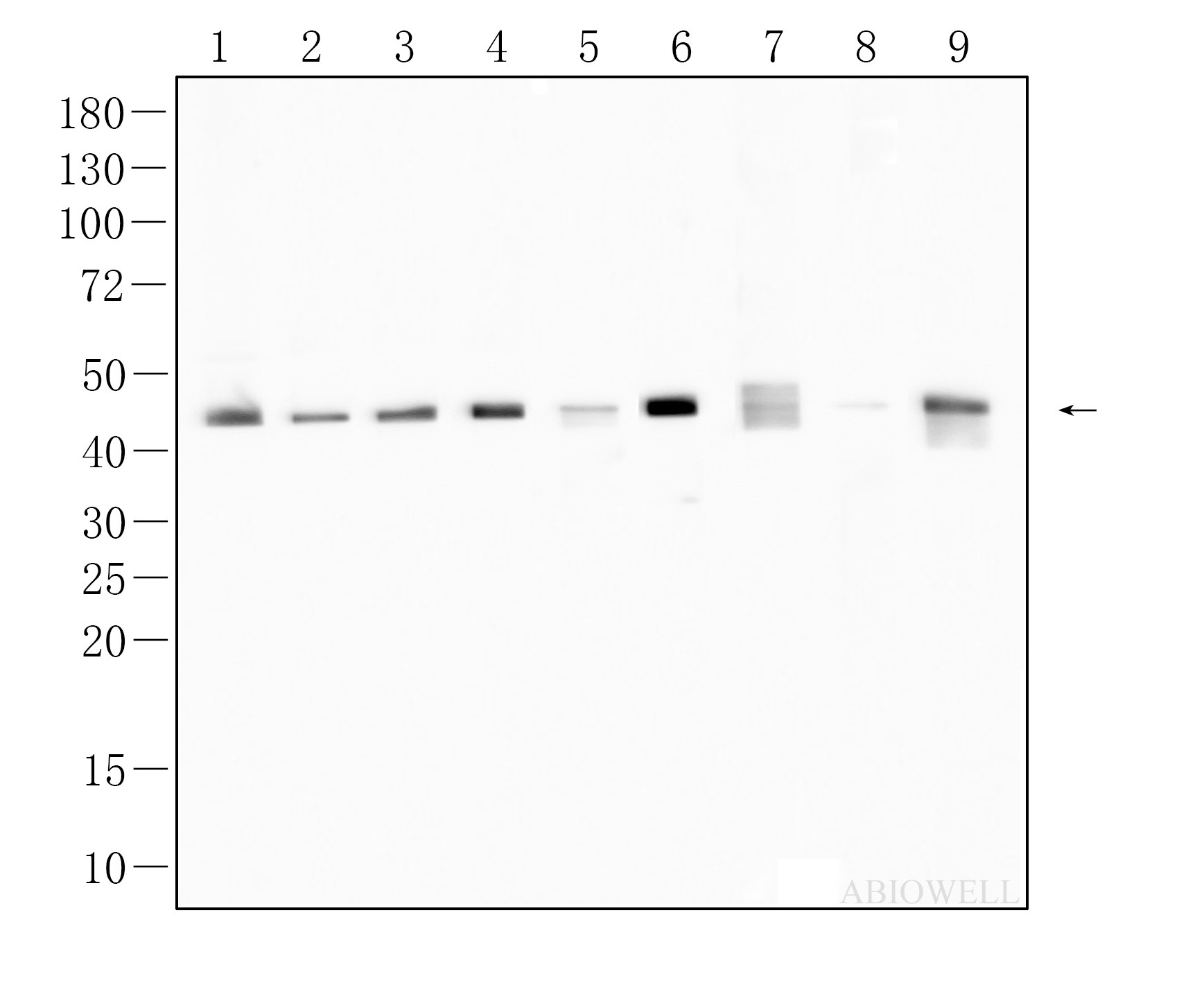
Fig : Western blot analysis of β-actin on different lysates. Proteins were transferred to a NC membrane and blocked with 5% NF-Milk in TBST for 1 hour at room temperature. The primary antibody (AWA80002, 1/10000) was used in TBST at room temperature for 2 hours. Goat Anti-Rabbit IgG - HRP Secondary Antibody (AWS0002) at 1:5,000 dilution was used for 1 hour at room temperature. Positive control: Lane 1: Hela cell Lane 2: K562 cell Lane 3: A549 cell Lane 4: COS7 cell Lane 5: PC-12 cell Lane 6: MCF-7 cell Lane 7: Raw264.7 cell Lane 8: THP-1 cell Exposure time: 45 seconds Predicted band size: 42 kDa Observed band size: 42 kDa |
1. Fan, Shuangshi et al. “Molecular prognostic of nine parthanatos death-related genes in glioma, particularly in COL8A1 identification.” Journal of neurochemistry vol. 168,3 (2024): 205-223. doi:10.1111/jnc.16049 . PubMed:38225203
2. Liu, Ying et al. “PPAR-α inhibits DHEA-induced ferroptosis in granulosa cells through upregulation of FADS2.” Biochemical and biophysical research communications vol. 715 (2024): 150005. doi:10.1016/j.bbrc.2024.150005. PubMed:38678785
3. Wang, Fan et al. “Silencing BMAL1 promotes M1/M2 polarization through the LDHA/lactate axis to promote GBM sensitivity to bevacizumab.” International immunopharmacology vol. 134 (2024): 112187. doi:10.1016/j.intimp.2024.112187. PubMed:38733825
4. Wu, Zhifeng et al. “Silencing p75NTR regulates osteogenic differentiation and angiogenesis of BMSCs to enhance bone healing in fractured rats.” Journal of orthopaedic surgery and research vol. 19,1 192. 20 Mar. 2024, doi:10.1186/s13018-024-04653-8. PubMed:38504358
5. Tian, Zhenyang et al. “LncARSR promotes glioma tumor growth by mediating glycolysis through the STAT3/HK2 axis.” Cytokine vol. 180 (2024): 156663. doi:10.1016/j.cyto.2024.156663. PubMed:38815522
-
-
- 50μL
- ¥580
- 1-3个工作日
-
- 100μL
- ¥920
- 1-3个工作日
-
- 500μL
- ¥3800
- 1-3个工作日
-
相关产品
-
Cdk6 Recombinant Rabbit Monoclonal Antibody
GAPDH Rabbit Polyclonal Antibody
GFAP Recombinant Mouse Monoclonal Antibody
Ki67 Rabbit Monoclonal Antibody
Stathmin 1 Recombinant Rabbit Monoclonal Antibody
HMGB1 Recombinant Rabbit Monoclonal Antibody
SQSTM1/p62 Mouse Monoclonal Antibody
p53 Recombinant Rabbit Monoclonal Antibody

