HDAC3 Recombinant Rabbit Monoclonal Antibody

-
-
- 20μL
- ¥620
- 1-3个工作日
-
- 50μL
- ¥1250
- 1-3个工作日
-
- 100μL
- ¥2200
- 1-3个工作日
Product Details
| Host Species: Rabbit | Reactivity: Human,Mouse,Rat | Molecular Wt: 49 kDa | |
Clonality: Monoclonal | Isotype: IgG | Concentration: 1 mg/ml | ||
Other Names: HDAC3; HDAC 3; HDAC3_HUMAN; Histone deacetylase 3; HD3; RPD3-2; RPD3 2; RPD3-2; SMAP45
| ||||
Formulation: Liquid in PBS containing 50% glycerol, 0.5% BSA and 0.02% sodium azide. | ||||
Purification: Affinity-chromatography | ||||
Storage: -20°C,1 year | ||||
Applications
| WB 1:1000-1:5000 IHC-P 1:50-1:1000 IF-C 1:50 IF-T 1:50 IP Use at an assay dependent concentration.
| |||
Immunogen Information | Gene Name: HDAC3 | Protein Name: Histone deacetylase 3 | ||
Gene ID: 8841 (Human) 15183 (Mouse) 84578 (Rat) | SwissPro: O15379 (Human) O88895 (Mouse) Q6P6W3 (Rat)
| |||
Subcellular Location: Nucleus. Cytoplasm. Cytoplasm, cytosol. | ||||
Immunogen: Synthetic peptide within C-terminal human HDAC3. | ||||
Specificity: HDAC3 Monoclonal Antibody detects endogenous levels of HDAC3 protein. | ||||
| Product images | |
|
|
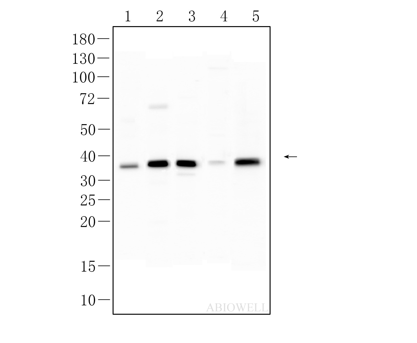
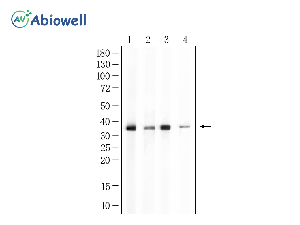
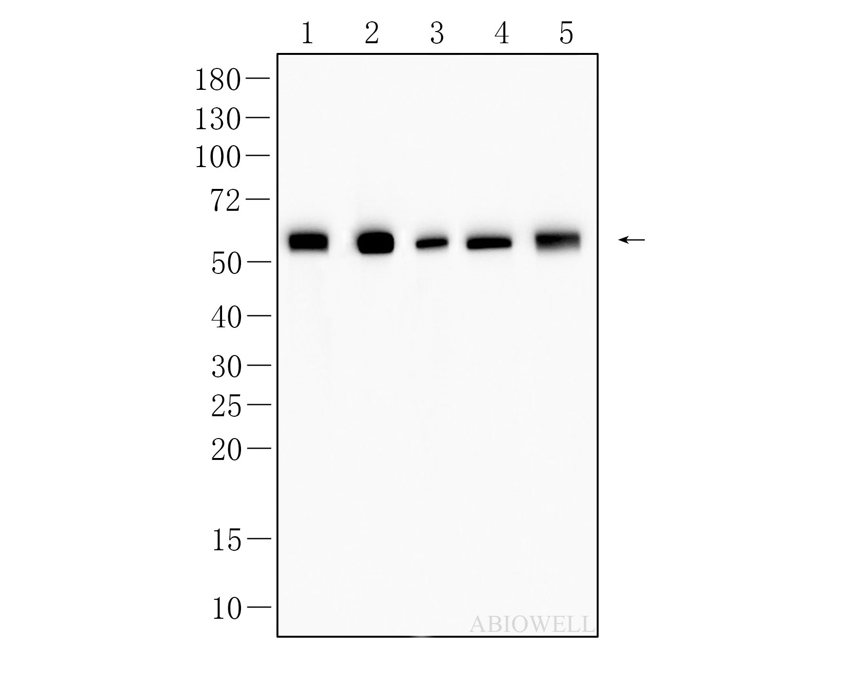
Fig : Western blot analysis of HDAC3 on different lysates. Proteins were transferred to a NC membrane and blocked with 5% NF-Milk in TBST for 1 hour at room temperature. The primary antibody (AWA12693, 1/1000) was used in TBST at room temperature for 2 hours. Goat Anti- Rabbit IgG - HRP Secondary Antibody (AWS0002) at 1:5,000 dilution was used for 1 hour at room temperature. Positive control: Lane 1: A431 cell Lane 2: HEK293 cell Lane 3: K562 cell Lane 4: HepG2 cell Lane 5: NIH/3T3 cell Lane 6: HEPA1-6 cell Lane 7: Mouse brain tissue Lane 8: Mouse testicular tissue Lane 9: Rat testicular tissue Predicted molecular weight:49KD Observed molecular weight:49KD Exposure time: 45 seconds |
|
|
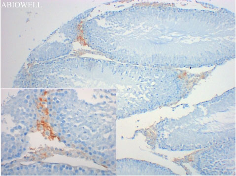
Fig : Immunohistochemical analysis of paraffin-embedded rat-testicle tissue with Rabbit anti-HDAC3 antibody ( AWA12693 ) at 1/200 dilution. The section was pre-treated using heat mediated antigen retrieval with Sodium citrate buffer (pH 6.0) for 20 minutes. The tissues were blocked in 3% H2O2 for 15 minutes at room temperature, washed with ddH2O and PBS, and then probed with the primary antibody ( AWA12693 ) at 1/200 dilution for 1 hour at room temperature. The detection was performed using an HRP conjugated compact polymer system(ABIOWELL, AWI0629). DAB was used as the chromogen. Tissues were counterstained with hematoxylin and mounted with DPX. |
|
|
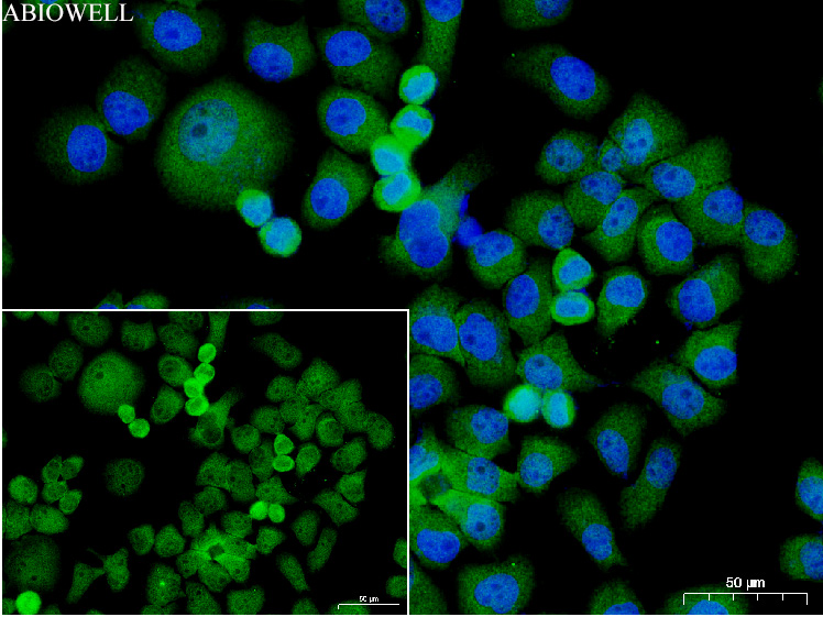
Fig: Immunocytochemistry analysis of Hela cells labeling HDAC3 with Rabbit anti-HDAC3 antibody (AWA12693)at 1/50 dilution(Green). Cells were fixed in 4% paraformaldehyde for 10 minutes at 37 ℃, permeabilized with 0.03% Triton X-100 in PBS for 30 minutes, and then blocked with 5% BSA for 60 minutes at 37 ℃. Cells were then incubated with Rabbit anti-HDAC3 antibody (AWA12693)at 1/50 dilution in 2% negative goat serum overnight at 4 ℃. Goat Anti-Rabbit IgG H&L (iFluor™ 488, AWS0005c) was used as the secondary antibody at 1/200 dilution for 60 minutes at 37 ℃. Nuclear DNA was labelled in blue with DAPI(AWC0291). |
|
|
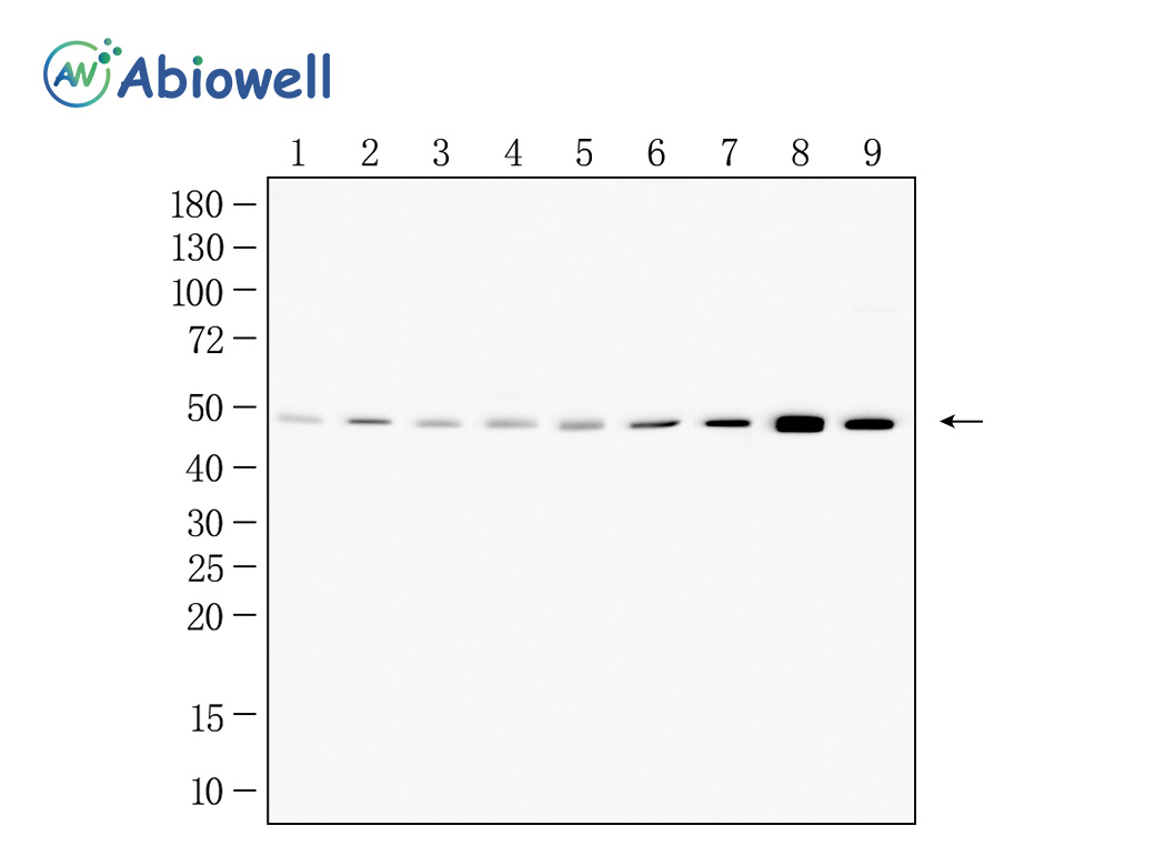
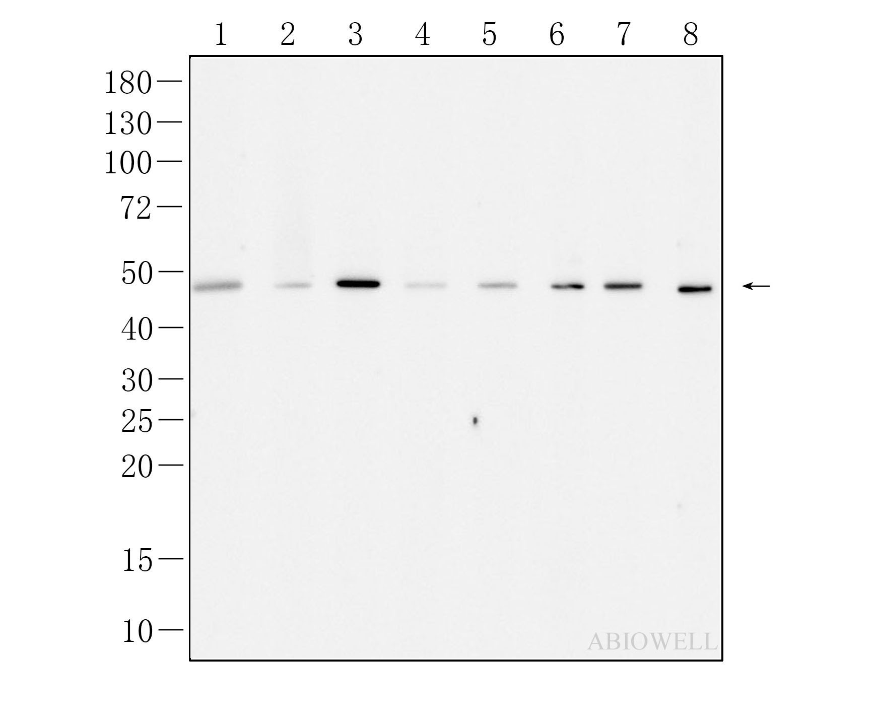
Fig : Western blot analysis of HDAC3 on different lysates. Proteins were transferred to a NC membrane and blocked with 5% NF-Milk in TBST for 1 hour at room temperature. The primary antibody (AWA12693, 1/1000) was used in TBST at room temperature for 2 hours. Goat Anti-rabbit IgG - HRP Secondary Antibody (AWS0002) at 1:5,000 dilution was used for 1 hour at room temperature. Positive control: Lane 1: RAW264.7 cell lysate Lane 2: Rat brain tissue lysate Lane 3: PC-12 cell lysate Lane 4: HUVEC cell lysate Lane 5: A549 cell lysate Lane 6: K562 cell lysate Lane 7: HeLa cell lysate Lane 8: NIH3T3 cell lysate |
|
|
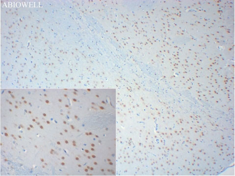
Fig : Immunohistochemical analysis of paraffin-embedded rat-brain tissue with Rabbit anti-HDAC3 antibody ( AWA12693 ) at 1/200 dilution. The section was pre-treated using heat mediated antigen retrieval with Sodium citrate buffer (pH 6.0) for 20 minutes. The tissues were blocked in 3% H2O2 for 15 minutes at room temperature, washed with ddH2O and PBS, and then probed with the primary antibody ( AWA12693 ) at 1/200 dilution for 1 hour at room temperature. The detection was performed using an HRP conjugated compact polymer system(ABIOWELL, AWI0629). DAB was used as the chromogen. Tissues were counterstained with hematoxylin and mounted with DPX. |
|
|
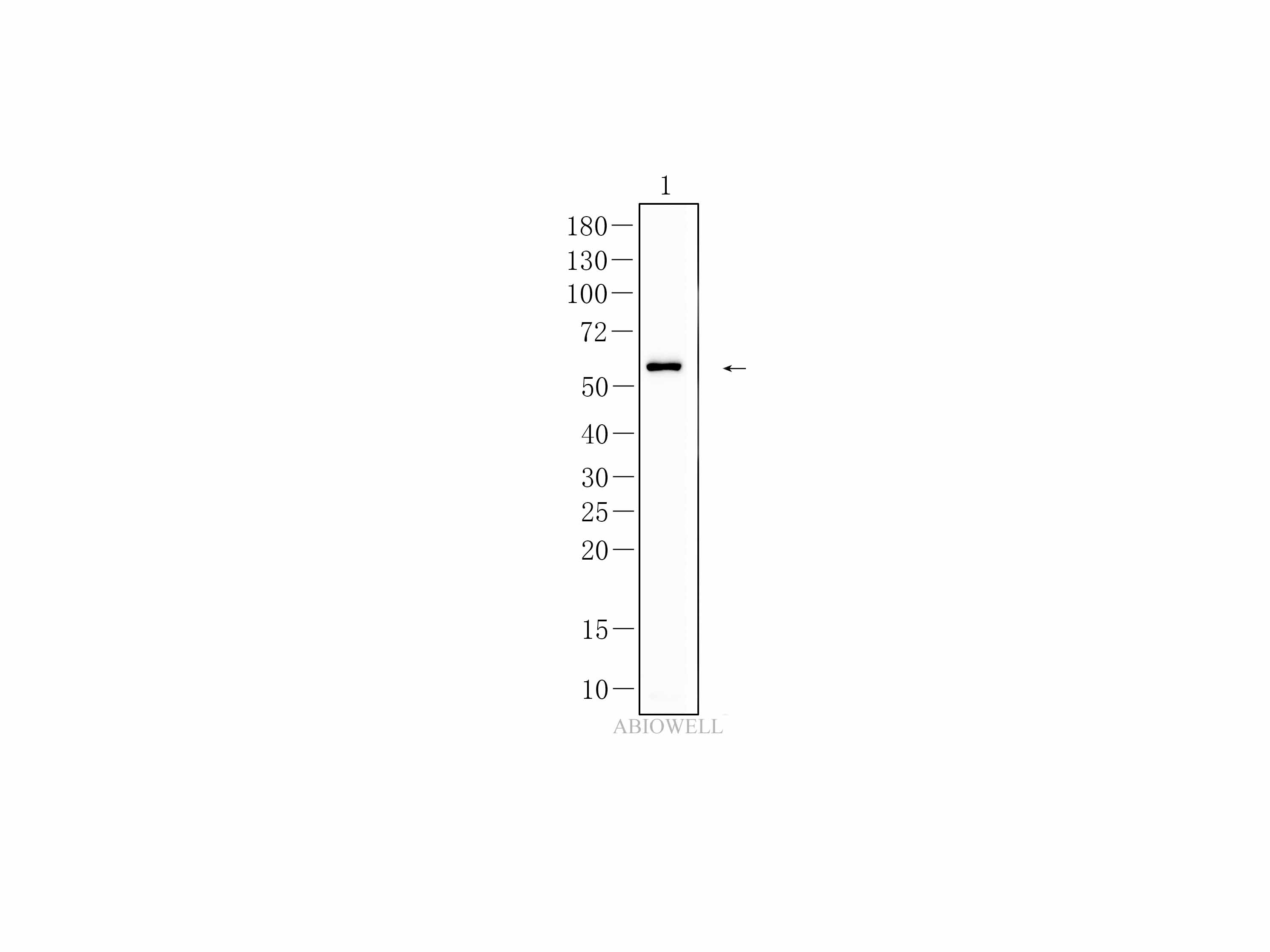
Fig : Western blot analysis of HDAC3 on different lysates. Proteins were transferred to a NC membrane and blocked with 5% NF-Milk in TBST for 1 hour at room temperature. The primary antibody (AWA12693, 1/1000) was used in TBST at room temperature for 2 hours. Goat Anti- Rabbit IgG - HRP Secondary Antibody (AWS0002) at 1:5,000 dilution was used for 1 hour at room temperature. Positive control: Lane 1: HL60 cell Lane 2: Raji cell Lane 3: RAW 264.7 cell Lane 4: PC12 cell Lane 5: HSC-T6 cell Lane 6: Rat brain tissue Predicted molecular weight:49KD Observed molecular weight:49KD Exposure time: 180 seconds |
-
-
- 20μL
- ¥620
- 1-3个工作日
-
- 50μL
- ¥1250
- 1-3个工作日
-
- 100μL
- ¥2200
- 1-3个工作日
-
相关产品
-
Cdk6 Recombinant Rabbit Monoclonal Antibody
GAPDH Rabbit Polyclonal Antibody
GFAP Recombinant Mouse Monoclonal Antibody
Ki67 Rabbit Monoclonal Antibody
Stathmin 1 Recombinant Rabbit Monoclonal Antibody
HMGB1 Recombinant Rabbit Monoclonal Antibody
SQSTM1/p62 Mouse Monoclonal Antibody
p53 Recombinant Rabbit Monoclonal Antibody