SQSTM1/p62 Recombinant Rabbit Monoclonal Antibody
-
-
- 20μL
- ¥620
- 1-3个工作日
-
- 50μL
- ¥1250
- 1-3个工作日
-
- 100μL
- ¥2200
- 1-3个工作日
|
Product Details |
Host Species: Rabbit |
Reactivity: Human, Mouse, Rat |
Molecular Wt: Predicted MW: 48 kDa | |||
|
|
Clonality: Monoclonal |
Isotype: IgG |
Concentration: 0.558mg/ml | |||
|
|
Other Names: P62; p60; p62B; EBIAP; FTDALS3; STAP; STONE14; ORCA; OSF-6; Osi; OSIL; ZIP 3; ZIP3; ZIP; Sequestosome 1; Sequestosome-1; PDB 3; PDB3; SQSTM 1; SQSTM1; Ubiquitin binding protein p62; SQSTM1/p62 | |||||
|
|
Formulation: Liquid in PBS containing 50% glycerol, 0.5% BSA and 0.02% sodium azide. | |||||
|
|
Purification: Affinity-chromatography | |||||
|
|
Storage: Store at -20°C. Stable for one year after shipment. Aliquoting is unnecessary for -20°C storage. | |||||
|
Applications |
WB 1:1000-1:10000 | |||||
|
Immunogen |
Gene Name: SQSTM1 |
Protein Name: Sequestosome-1 | ||||
|
|
Gene ID: 8878 (Human) |
SwissPro: Q13501 (Human) | ||||
|
Immunogen |
Subcellular Location: Cytoplasmic vesicle, autophagosome. Preautophagosomal structure. Cytoplasm, cytosol. Nucleus, PML body. Late endosome. Lysosome. Nucleus. Endoplasmic reticulum. Cytoplasm, myofibril, sarcomere. | |||||
|
|
Immunogen: Synthetic peptide within human SQSTM1/p62. AA range: 400 to the C-terminus. | |||||
|
|
Specificity: SQSTM1/p62 Monoclonal Antibody detects endogenous levels of SQSTM1/p62 protein. | |||||
| Product images | |
|
Fig:Flow cytometric analysis of HELA cells labeling SQSTM1/P62. Overlay histogram showing HELA cells stained with SQSTM1/P62 (green line). The cell were fixed in 4% paraformaldehyde for 30 minutes at 37 ℃, permeabilized with 0.02% Triton X-100 in PBS for 30 minutes,and then stained with the primary antibody(AWA10515, 1:50 ) for 30 min at 4°C. The secondary antibody used was an Alexa Fluor 488-conjugated Goat anti-Rabbit IgG Secondary antibody (AWS0005b) at 1/1000 dilution for 30 min at 4ºC. Unlabelled sample was used as a control (cells without incubation with primary antibody; red). |
|
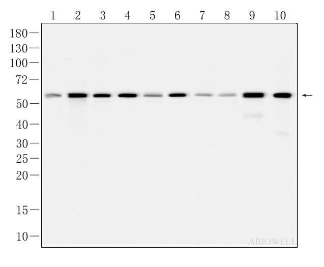
Fig : Western blot analysis of SQSTM1/p62 on different lysates. Proteins were transferred to a NC membrane and blocked with 5% NF-Milk in TBST for 90 minutes at room temperature. The primary antibody (AWA10515, 1/1000) was used in TBST at room temperature for 90 minutes. Goat Anti-Rabbit IgG - HRP Secondary Antibody (AWS0002) at 1:5,000 dilution was used for 1 hour at room temperature. Positive control: Lane 1: SHZ-88 cell Lane 2: RBL-2H3 cell Lane 3: HeLa cell Lane 4: NRF-49F cell Lane 5: NIH3T3 cell Lane 6: HEK293 cell Lane 7: HepG2 cell Lane 8: HCT116 cell Predicted molecular weight: 48 kDa Observed molecular weight: 62 kDa |
|
Fig: Immunocytochemistry analysis of Hela cells labeling SQSTM1/p62 with Rabbit anti-SQSTM1/p62 antibody (AWA10515) at 1/300 dilution(Green). Cells were fixed in 4% paraformaldehyde for 10 minutes at 37 ℃, permeabilized with 0.03% Triton X-100 in PBS for 30 minutes, and then blocked with 5% BSA for 60 minutes at 37 ℃. Cells were then incubated with Rabbit anti-SQSTM1/p62 antibody (AWA10515)at 1/300 dilution in 2% negative goat serum overnight at 4 ℃. Goat Anti-Rabbit IgG H&L (iFluor™ 488, AWS0005c) was used as the secondary antibody at 1/200 dilution for 60 minutes at 37 ℃. Nuclear DNA was labelled in blue with DAPI(AWC0291). |
|
Fig : Western blot analysis of SQSTM1/p62 on different lysates. Proteins were transferred to a NC membrane and blocked with 5% NF-Milk in TBST for 1 hour at room temperature. The primary antibody (AWA10515, 1/1000) was used in TBST at room temperature for 2 hours. Goat Anti-Rabbit IgG - HRP Secondary Antibody (AWS0002) at 1:5,000 dilution was used for 1 hour at room temperature. Positive control: Lane 1: U87-MG cell Lane 2: SK-BR-3 cell Lane 3: CTX cell Lane 4: PANC-1 cell Predicted molecular weight:48 kDa Observed molecular weight:62 kDa |
|
Fig : Immunohistochemical analysis of paraffin-embedded Rat-spleen tissue with Rabbit anti-SQSTM1/P62 antibody (AWA10515) at 1/200 dilution. The section was pre-treated using heat mediated antigen retrieval with Sodium citrate buffer (pH 6.0) for 20 minutes. The tissues were blocked in 3% H2O2 for 15 minutes at room temperature, washed with ddH2O and PBS, and then probed with the primary antibody ( AWA10515 ) at 1/200 dilution for 1 hour at room temperature. The detection was performed using an HRP conjugated compact polymer system(ABIOWELL, AWI0629). DAB was used as the chromogen. Tissues were counterstained with hematoxylin and mounted with DPX. |
|
Fig : Immunohistochemical analysis of paraffin-embedded Rat-colon tissue with Rabbit anti-SQSTM1/P62 antibody (AWA10515) at 1/200 dilution. The section was pre-treated using heat mediated antigen retrieval with Sodium citrate buffer (pH 6.0) for 20 minutes. The tissues were blocked in 3% H2O2 for 15 minutes at room temperature, washed with ddH2O and PBS, and then probed with the primary antibody ( AWA10515 ) at 1/200 dilution for 1 hour at room temperature. The detection was performed using an HRP conjugated compact polymer system(ABIOWELL, AWI0629). DAB was used as the chromogen. Tissues were counterstained with hematoxylin and mounted with DPX. |
|
Fig: Immunocytochemistry analysis of U-251MG cells labeling SQSTM1/p62 with Rabbit anti-SQSTM1/p62 antibody (AWA10515) at 1/100 dilution(green). Cells were fixed in 4% paraformaldehyde for 20 minutes at 37 ℃, permeabilized with 0.03% Triton X-100 in PBS for 30 minutes, and then blocked with 5% BSA for 60 minutes at 37 ℃. Cells were then incubated with rabbit anti-P62 antibody (AWA10515) at 1/100 dilution in 2% negative goat serum overnight at 4 ℃. Goat Anti-rabbit IgG H&L (iFluor™ 488, AWS0005) was used as the secondary antibody at 1/200 dilution for 60 minutes at 37 ℃. Nuclear DNA was labelled in blue with DAPI(AWC0291). |
|
Fig: Fluorescence immunohistochemical analysis of Rat-cerebral cortex tissue (Formalin/PFA-fixed paraffin-embedded sections). with Rabbit anti-SQSTM1/p62 antibody (AWA10515) at 1/200 dilution. The immunostaining was performed with the TSA Immuno-staining Kit (ABIOWELL, AWI0688). The section was pre-treated using heat mediated antigen retrieval with Sodium citrate buffer (pH 6.0) for 20 minutes. The tissues were blocked in 3% H2O2 for 15 minutes at room temperature, washed with ddH2O and PBS, and then probed with the primary antibody (AWA10515) at 1/200 dilution for 1 hour at room temperature. The detection was performed using an HRP conjugated compact polymer system followed by a separate fluorescent tyramide signal amplification system (green). DAPI (blue, AWC0291) was used as a nuclear counter stain. Image acquisition was performed with Slide Scanner. |
|
Fig:Immunoprecipitation of SQSTM1 from U87-MG cells was performed using SQSTM1 Rabbit mAb (AWA10515,1:250). Rabbit IgG isotype control was used to precipitate the Control IgG sample. The IP sample was eluted with Glycine buffer. Western blot analysis of immunoprecipitates was conducted using SQSTM1 Rabbit mAb (AWA10515) at a dilution of 1:1000. Goat Anti-Rabbit IgG(H+L) - HRP Secondary Antibody (AWS0002) at 1:5,000 dilution. |
引用文献 (1)
Ethnopharmacological relevance WuHuTang (WHT) is a traditional Chinese medicine compound for treating asthma, and the evidence supports that it has a good effect on acute asthma attacks in children and adults. Respiratory syncytial virus (RSV) is an important factor in the pathogenesis of acute asthma attacks, and the effect on dendritic cells is the key to its pathogenesis. Previous studies have confirmed that the pathogenesis of viruses is related to exosomes. However, there are few studies on the exosomes induced by RSV. Whether WHT can improve the changes caused by RSV-induced exosomes or not is worthy of further exploration. Aim of the study We aim to study the effects of RSV-induced exosomes on the function and autophagy of dendritic cells, and to observe the intervention effect of WHT serum on the above effects. Methods The co-culture model of exosomes derived from bone marrow mesenchymal stem cells induced by RSV (BMSCs-Exo-RSV) and dendritic cells was established, and then WHT serum was used to intervene. After 24 h of intervention, the CCK-8 method, flow cytometry, Elisa, RT-qCPR, and Western blot were used to detect the above-mentioned culture model. Results RSV-induced exosomes had certain effects on viability, apoptosis, and costimulatory molecules generation of dendritic cells. At the same time, the levels of IL-6, IL-12, TNF-α, and autophagy increased, while the levels of IL-4, IL-10, and TGF-β decreased, and the AKT/TSC/mTOR pathway was inhibited. WHT serum could activate this pathway and reverse the above changes in dendritic cells. Conclusion This study reveals that the pathogenic effect of RSV is related to the exosomes induced by RSV. The exosomes induced by RSV affect the function of dendritic cells by inhibiting the AKT/TSC/mTOR pathway, which can be activated by WHT to reverse the effects caused by RSV-induced exosomes.
-
-
- 20μL
- ¥620
- 1-3个工作日
-
- 50μL
- ¥1250
- 1-3个工作日
-
- 100μL
- ¥2200
- 1-3个工作日
-
相关产品
-
CD31 Recombinant Rabbit Monoclonal Antibody
Occludin Recombinant Rabbit Monoclonal Antibody
Occludin Recombinant Mouse Monoclonal Antibody
O-Linked N-Acetylglucosamine Recombinant Mouse Monoclonal Antibody
Biotin Recombinant Rabbit Monoclonal Antibody
Biotin Recombinant Mouse Monoclonal Antibody
RUNX2 Recombinant Mouse Monoclonal Antibody
KiSS-1R Recombinant Rabbit Monoclonal Antibody