Vimentin Recombinant Rabbit Monoclonal Antibody


-
-
- 20μL
- ¥620
- 有库存
-
- 50μL
- ¥1250
- 有库存
-
- 100μL
- ¥2200
- 有库存
Product Details
| Host Species: Rabbit | Reactivity: Human,Mouse,Rat | Molecular Wt: 54 kDa | |
Clonality: Monoclonal | Isotype: IgG | Concentration: 1 mg/ml | ||
Other Names: VIM; Vimentin | ||||
Formulation: Liquid in PBS containing 50% glycerol, 0.5% BSA and 0.02% sodium azide. | ||||
Purification: Affinity-chromatography | ||||
Storage: -20°C,1 year | ||||
Applications
| WB 1:20000 IHC-P 1:100-1:10000 IHC-F 1:50 IF 1:100-1:500 FC 1:50-1:100 IP Use at an assay dependent concentration.
| |||
Immunogen Information | Gene Name: VIM | Protein Name: Vimentin | ||
Gene ID: 7431 (Human) 22352 (Mouse) 81818 (Rat) | SwissPro: P08670 (Human) P20152 (Mouse) P31000 (Rat)
| |||
Immunogen: Synthetic peptide within C-terminal human Vimentin. | ||||
Specificity: Vimentin Monoclonal Antibody detects endogenous levels of Vimentin protein. | ||||
| Product images | |
|
|
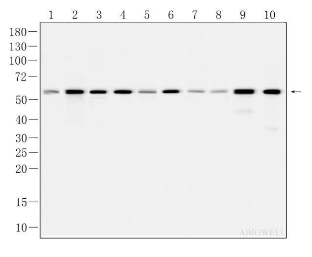
Fig : Western blot analysis of Vimentin on different lysates. Proteins were transferred to a NC membrane and blocked with 5% NF-Milk in TBST for 1 hour at room temperature. The primary antibody (AWA10146, 1/1000) was used in TBST at room temperature for 2 hours. Goat Anti-Rabbit IgG - HRP Secondary Antibody (AWS0002) at 1:5,000 dilution was used for 1 hour at room temperature. Positive control: Lane 1: HEK293 cell Lane 2: Jurkat cell Lane 3: A549 cell Lane 4: NIH/3T3 cell Lane 5: RBL-2H3 cell Lane 6: NRK-49F cell Lane 7: PC12 cell Lane 8: RAW264.7 cell Predicted molecular weight:54KD Observed molecular weight:54KD |
|
|
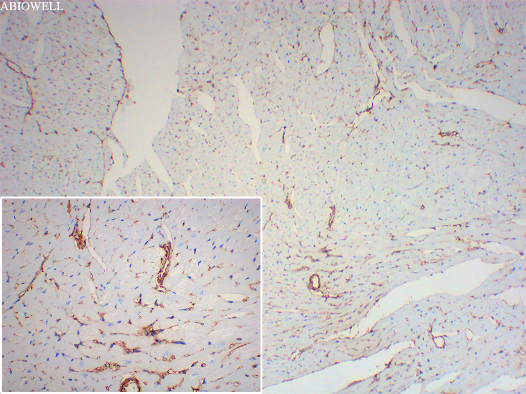
Fig : Immunohistochemical analysis of paraffin-embedded Mouse-heart tissue with Rabbit anti-vimentin (AWA10146) at 1/800 dilution. The section was pre-treated using heat mediated antigen retrieval with Sodium citrate buffer (pH 6.0) for 20 minutes. The tissues were blocked in 3% H2O2 for 15 minutes at room temperature, washed with ddH2O and PBS, and then probed with the primary antibody (AWA10146) at 1/800 dilution for 1 hour at room temperature. The detection was performed using an HRP conjugated compact polymer system(ABIOWELL, AWI0629). DAB was used as the chromogen. Tissues were counterstained with hematoxylin and mounted with DPX. |
|
|
Fig : Immunohistochemical analysis of paraffin-embedded Mouse-kidney tissue with Rabbit anti-Vimentin (AWA10146) at 1/200 dilution. The section was pre-treated using heat mediated antigen retrieval with Sodium citrate buffer (pH 6.0) for 20 minutes. The tissues were blocked in 3% H2O2 for 15 minutes at room temperature, washed with ddH2O and PBS, and then probed with the primary antibody (AWA10146) at 1/200 dilution for 1 hour at room temperature. The detection was performed using an HRP conjugated compact polymer system(ABIOWELL, AWI0629). DAB was used as the chromogen. Tissues were counterstained with hematoxylin and mounted with DPX. |
|
|
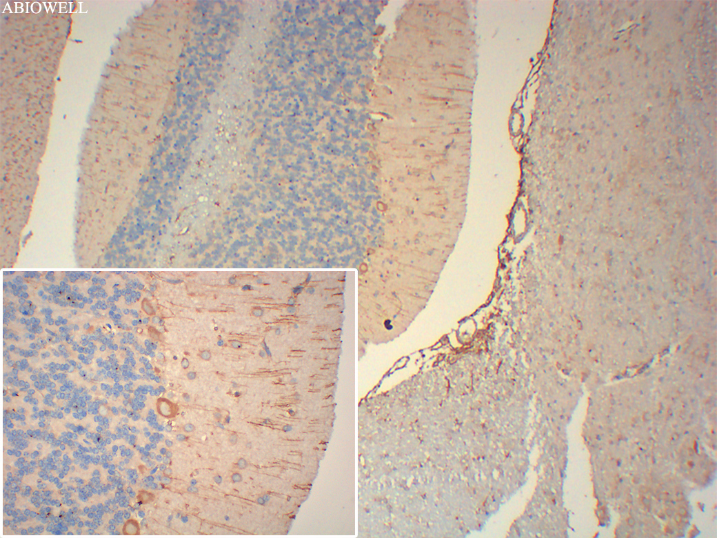
Fig : Immunohistochemical analysis of paraffin-embedded Rat-cerebellum tissue with Rabbit anti-Vimentin (AWA10146) at 1/200 dilution. The section was pre-treated using heat mediated antigen retrieval with Sodium citrate buffer (pH 6.0) for 20 minutes. The tissues were blocked in 3% H2O2 for 15 minutes at room temperature, washed with ddH2O and PBS, and then probed with the primary antibody (AWA10146) at 1/200 dilution for 1 hour at room temperature. The detection was performed using an HRP conjugated compact polymer system(ABIOWELL, AWI0629). DAB was used as the chromogen. Tissues were counterstained with hematoxylin and mounted with DPX. |
|
|
Fig : Immunohistochemical analysis of paraffin-embedded Rat-heart tissue with Rabbit anti-vimentin (AWA10146) at 1/800 dilution. The section was pre-treated using heat mediated antigen retrieval with Sodium citrate buffer (pH 6.0) for 20 minutes. The tissues were blocked in 3% H2O2 for 15 minutes at room temperature, washed with ddH2O and PBS, and then probed with the primary antibody (AWA10146) at 1/800 dilution for 1 hour at room temperature. The detection was performed using an HRP conjugated compact polymer system(ABIOWELL, AWI0629). DAB was used as the chromogen. Tissues were counterstained with hematoxylin and mounted with DPX. |
|
|
Fig : Immunohistochemical analysis of paraffin-embedded Rat-myocardium tissue with Rabbit anti-Vimentin (AWA10146) at 1/200 dilution. The section was pre-treated using heat mediated antigen retrieval with Sodium citrate buffer (pH 6.0) for 20 minutes. The tissues were blocked in 3% H2O2 for 15 minutes at room temperature, washed with ddH2O and PBS, and then probed with the primary antibody (AWA10146) at 1/200 dilution for 1 hour at room temperature. The detection was performed using an HRP conjugated compact polymer system(ABIOWELL, AWI0629). DAB was used as the chromogen. Tissues were counterstained with hematoxylin and mounted with DPX. |
|
|
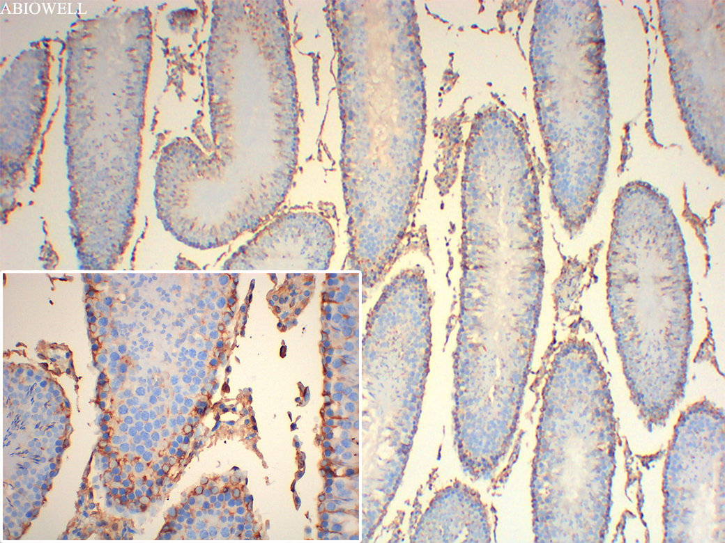
Fig : Immunohistochemical analysis of paraffin-embedded Rat-testis tissue with Rabbit anti-Vimentin (AWA10146) at 1/200 dilution. The section was pre-treated using heat mediated antigen retrieval with Sodium citrate buffer (pH 6.0) for 20 minutes. The tissues were blocked in 3% H2O2 for 15 minutes at room temperature, washed with ddH2O and PBS, and then probed with the primary antibody (AWA10146) at 1/200 dilution for 1 hour at room temperature. The detection was performed using an HRP conjugated compact polymer system(ABIOWELL, AWI0629). DAB was used as the chromogen. Tissues were counterstained with hematoxylin and mounted with DPX. |
-
-
- 20μL
- ¥620
- 1-3个工作日
-
- 50μL
- ¥1250
- 1-3个工作日
-
- 100μL
- ¥2200
- 1-3个工作日
-
相关产品
-
Cytokeratin 15 Recombinant Rabbit Monoclonal Antibody
HMGB1 Recombinant Rabbit Monoclonal Antibody
SQSTM1/p62 Mouse Monoclonal Antibody
Vimentin Mouse Monoclonal Antibody
xCT/SLC7A11 Recombinant Rabbit Monoclonal Antibody
SQSTM1/p62 Recombinant Rabbit Monoclonal Antibody
Caspase-8 Recombinant Rabbit Monoclonal Antibody
Ki67 Recombinant Rabbit Monoclonal Antibody