NFkB p65 Mouse Monoclonal Antibody


-
-
- 20μL
- ¥620
- 有库存
-
- 50μL
- ¥1250
- 有库存
-
- 100μL
- ¥2200
- 有库存
Product Details
| Host Species: Mouse | Reactivity: Human,Mouse,Rat | Molecular Wt: 65 kDa | |
Clonality: Monoclonal | ||||
Other Names: RELA; NFKB3; Transcription factor p65; Nuclear factor NF-kappa-B p65 subunit; Nuclear factor of kappa light polypeptide gene enhancer in B-cells 3
| ||||
Formulation: Liquid in PBS containing 50% glycerol, 0.5% BSA and 0.02% sodium azide. | ||||
Purification: Affinity-chromatography | ||||
Storage: -20°C,1 year | ||||
Applications
| WB 1:1000-1:3000 IHC 1:50-1:300 IF 1:200 IP 1:200
| |||
Immunogen Information | Gene Name: RELA | Protein Name: Transcription factor p65 | ||
Gene ID: 5970 (Human) 19697 (Mouse) 309165 (Rat) | SwissPro: Q04206 (Human) Q04207 (Mouse)
| |||
Immunogen: Recombinant Protein of human Transcription factor p65 AA range: 200-300. | ||||
Specificity: NFkB p65 Monoclonal Antibody detects endogenous levels of NFkB p65 protein. | ||||
| Product images | |
|
|
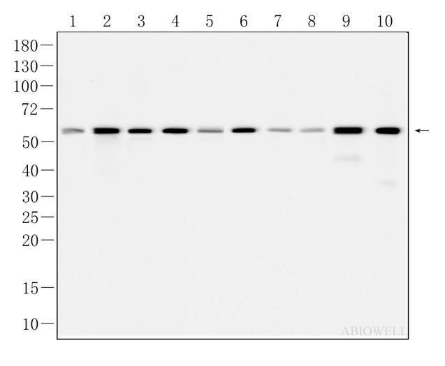
Fig : Western blot analysis of NF-Kβ p65 on different lysates. Proteins were transferred to a NC membrane and blocked with 5% NF-Milk in TBST for 1 hour at room temperature. The primary antibody (AWA00848, 1/1000) was used in TBST at room temperature for 2 hours. Goat Anti-Rabbit IgG - HRP Secondary Antibody (AWS0002) at 1:5,000 dilution was used for 1 hour at room temperature. Positive control: Lane 1: HELA cell Lane 2: HEK-293 cell Lane 3: Jurkat cell Lane 4: RAW264.7 cell Lane 5: NIH3T3 cell |
|
|
Fig: Immunocytochemistry analysis of NIH/3T3 cells labeling NFkB P65 with Mouse anti-NFkB P65 antibody (AWA00848)at 1/50 dilution(Red). Cells were fixed in 4% paraformaldehyde for 10 minutes at 37 ℃, permeabilized with 0.03% Triton X-100 in PBS for 30 minutes, and then blocked with 5% BSA for 60 minutes at 37 ℃. Cells were then incubated with Mouse anti-NFkB P65 antibody (AWA00848)at 1/50 dilution in 2% negative goat serum overnight at 4 ℃. Goat Anti-Mouse IgG H&L (iFluor™ 567, AWS0004c) was used as the secondary antibody at 1/200 dilution for 60 minutes at 37 ℃. Nuclear DNA was labelled in blue with DAPI(AWC0291). |
-
-
- 20μL
- ¥620
- 1-3个工作日
-
- 50μL
- ¥1250
- 1-3个工作日
-
- 100μL
- ¥2200
- 1-3个工作日
-
相关产品
-
Cytokeratin 15 Recombinant Rabbit Monoclonal Antibody
HMGB1 Recombinant Rabbit Monoclonal Antibody
SQSTM1/p62 Mouse Monoclonal Antibody
Vimentin Mouse Monoclonal Antibody
xCT/SLC7A11 Recombinant Rabbit Monoclonal Antibody
SQSTM1/p62 Recombinant Rabbit Monoclonal Antibody
Caspase-8 Recombinant Rabbit Monoclonal Antibody
Ki67 Recombinant Rabbit Monoclonal Antibody