
CD71 Rabbit Polyclonal Antibody
-
-
- 20μL
- ¥620
- 1-3个工作日
-
- 50μL
- ¥1250
- 1-3个工作日
-
- 100μL
- ¥2200
- 1-3个工作日
Product Details | Host Species: Rabbit | Reactivity: Human,Mouse | Molecular Wt: 85 kDa | |
Clonality: Polyclonal | Isotype: IgG | Concentration: 1mg/ml | ||
Other Names: TFRC; Transferrin receptor protein 1; TR; TfR; TfR1; Trfr; T9; p90; CD antigen CD71 | ||||
Formulation: Liquid in PBS containing 50% glycerol, 0.5% BSA and 0.02% sodium azide. | ||||
Purification: Affinity-chromatography | ||||
Storage: Store at -20°C. Stable for one year after shipment. Aliquoting is unnecessary for -20°C storage. | ||||
Applications | WB 1:500-1:2000 | |||
Immunogen Information | Gene Name: TFRC | Protein Name: Transferrin receptor protein 1 | ||
Gene ID: 7037 (Human) | SwissPro: P02786 (Human) | |||
Subcellular Location: Cell membrane. Melanosome. Secreted. | ||||
Immunogen: Recombinant protein within human CD71. AA range: 463-750. | ||||
Specificity: CD71 Polyclonal Antibody detects endogenous levels of CD71 protein. | ||||
| Product images | |
|
|
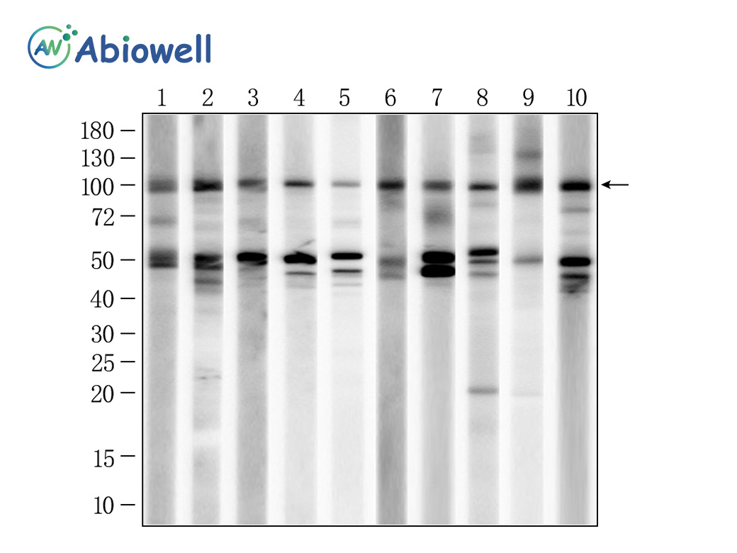
Fig : Western blot analysis of CD71 on different lysates. Proteins were transferred to a NC membrane and blocked with 5% NF-Milk in TBST for 1 hour at room temperature. The primary antibody ( AWA58276, 1/1000) was used in TBST(0.3%TWEEN20) at room temperature for 2 hours. Goat Anti-Rabbit IgG - HRP Secondary Antibody (AWS0002) at 1:5,000 dilution was used for 1 hour at room temperature.(10% SDS-PAGE gel.) Positive control: Lane 1: A549 cell Lane 2: Hela cell Lane 3: Jurkat cell Lane 4: K562 cell Lane 5: Raji cell Lane 6: HUVEC cell Lane 7: HT1080 cell Lane 8: PC-3 cell Lane 9: MCF-7 cell Lane 10: U87-MG cell Predicted molecular weight: 85 kDa Observed molecular weight: 100 kDa Exposure time: 90 seconds |
|
|
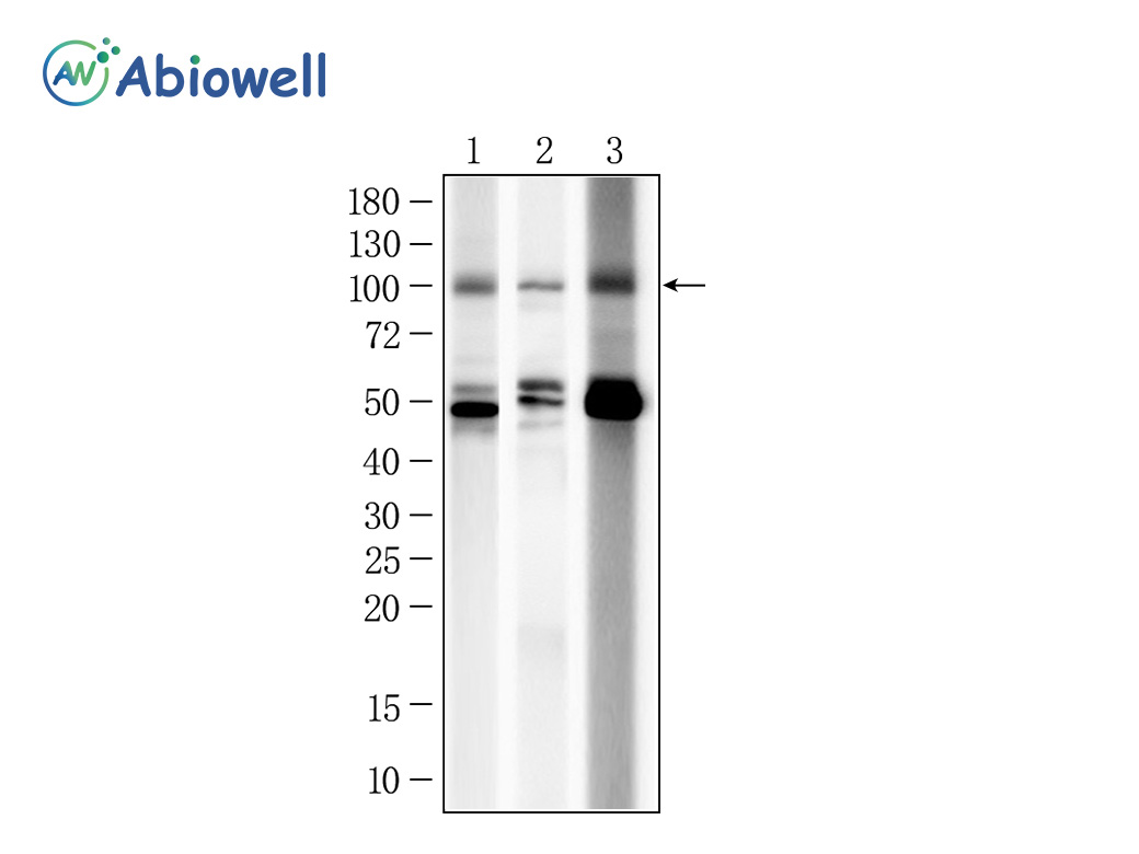
Fig : Western blot analysis of CD71 on different lysates. Proteins were transferred to a NC membrane and blocked with 5% NF-Milk in TBST for 1 hour at room temperature. The primary antibody ( AWA58276, 1/1000) was used in TBST(0.3%TWEEN20) at room temperature for 2 hours. Goat Anti-Rabbit IgG - HRP Secondary Antibody (AWS0002) at 1:5,000 dilution was used for 1 hour at room temperature.(10% SDS-PAGE gel.) Positive control: Lane 1: SW480 cell Lane 2: HaCaT cell Lane 3: HepG2 cell Predicted molecular weight: 85 kDa Observed molecular weight: 100 kDa Exposure time: 90 seconds |
|
|
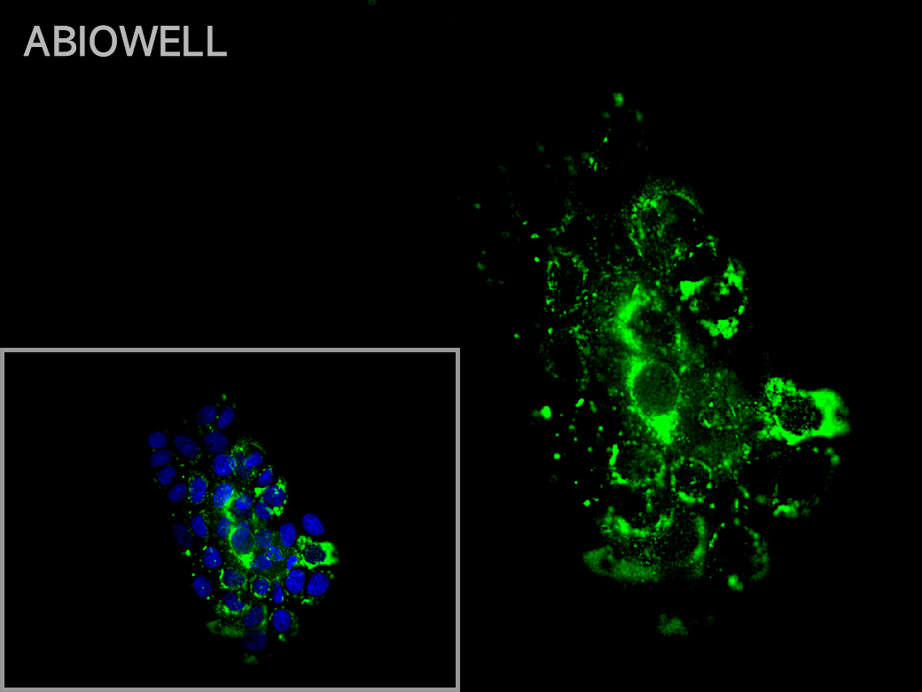
Fig: Immunocytochemistry analysis of A431 cells labeling CD71 with Rabbit anti-CD71 antibody (AWA58276) at 1/50 dilution(green). Cells were fixed in 4% paraformaldehyde for 10 minutes at 37 ℃, permeabilized with 0.03% Triton X-100 in PBS for 30 minutes, and then blocked with 5% BSA for 60 minutes at 37 ℃. Cells were then incubated with Rabbit anti-CD71 antibody (AWA58276) at 1/50 dilution in 2% negative goat serum overnight at 4 ℃. Goat Anti-Rabbit IgG H&L (iFluor™ 488, AWS0005) was used as the secondary antibody at 1/200 dilution for 60 minutes at 37 ℃. Nuclear DNA was labelled in blue with DAPI(AWC0291). |
-
-
- 20μL
- ¥620
- 1-3个工作日
-
- 50μL
- ¥1250
- 1-3个工作日
-
- 100μL
- ¥2200
- 1-3个工作日
-
相关产品
-
Cdk6 Recombinant Rabbit Monoclonal Antibody
GAPDH Rabbit Polyclonal Antibody
GFAP Recombinant Mouse Monoclonal Antibody
Ki67 Rabbit Monoclonal Antibody
HMGB1 Recombinant Rabbit Monoclonal Antibody
SQSTM1/p62 Mouse Monoclonal Antibody
Bcl-2 Recombinant Rabbit Monoclonal Antibody
SOD2 Rabbit Polyclonal Antibody


