×

1 / 1

xCT/SLC7A11 Recombinant Mouse Monoclonal Antibody [M43D07]
 -

一键复制产品信息

货号:
AWA00502
应用:
WB,IHC-P,IF-C,FCM
反应性:
Human,Mouse,Rat
来源:
Mouse
  • 20μL
  • ¥620
  • 1-3个工作日
  • 50μL
  • ¥1250
  • 1-3个工作日
  • 100μL
  • ¥2200
  • 1-3个工作日
  • 产品概述
  • Product Details

    Host Species:

    Mouse

    Reactivity:

    Human, Mouse, Rat

    Molecular Wt:

    Predicted MW: 55 kDa
    Observed MW: 35-40, 55 kDa


    Clonality:

    Monoclonal

    Isotype:

    IgG2a

    Concentration:

    0.578mg/ml


    Other Names:

    CCBR1; Cysteine/glutamate transporter; Calcium channel blocker resistance protein CCBR1; SLC7A11; Solute carrier family 7 member 11; xCT; Amino acid transport system xc-; Amino acid transport system xc xCT; xCT/SLC7A11


    Formulation:

    Liquid in PBS containing 50% glycerol, 0.5% BSA and 0.02% sodium azide.


    Purification:

    Affinity-chromatography


    Storage:

    Store at -20°C. Stable for one year after shipment. Aliquoting is unnecessary for -20°C storage.

    Applications

    WB 1:1000-1:5000
    IHC-P 1:100-1:1000
    IF-C 1:100-1:800
    FCM 1:50-1:200

    Immunogen
    Information

    Gene Name:

    SLC7A11

    Protein Name:

    Cystine/glutamate transporter


    Gene ID:

    23657 (Human)
    26570 (Mouse)

    SwissPro:

    Q9UPY5 (Human)
    Q9WTR6 (Mouse)

    Immunogen
    Information

    Subcellular Location:

    Cell membrane. Lysosome membrane. Cell projection, microvillus membrane.


    Immunogen:

    Recombinant protein within human xCT/SLC7A11.


    Specificity:

    xCT/SLC7A11 Monoclonal Antibody detects endogenous levels of xCT/SLC7A11 protein.


    Product images
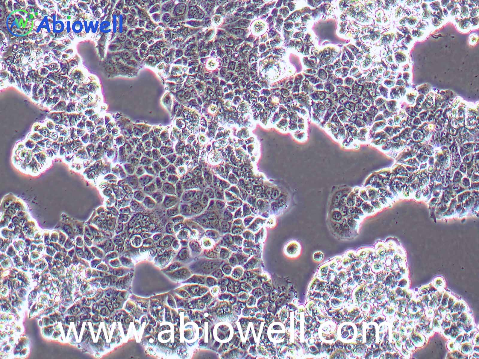
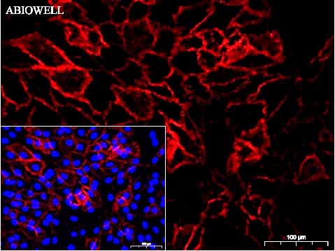
    xCT/SLC7A11 Recombinant Mouse Monoclonal Antibody [M43D07] - 1 Fig: Immunocytochemistry analysis of A549 cells labeling xCT/SLC7A11 with mouse anti-xCT/SLC7A11 antibody (AWA00502) at 1/100 dilution(Red).
    Cells were fixed in 4% paraformaldehyde for 10 minutes at 37 ℃, permeabilized with 0.03% Triton X-100 in PBS for 30 minutes, and then blocked with 5% BSA for 60 minutes at 37 ℃. Cells were then incubated with mouse anti-xCT/SLC7A11 antibody (AWA00502) at 1/100 dilution in 2% negative goat serum overnight at 4 ℃. Goat Anti-mouse IgG H&L (iFluor™ 594, AWS0004) was used as the secondary antibody at 1/200 dilution for 60 minutes at 37 ℃. Nuclear DNA was labelled in blue with DAPI(AWC0291).
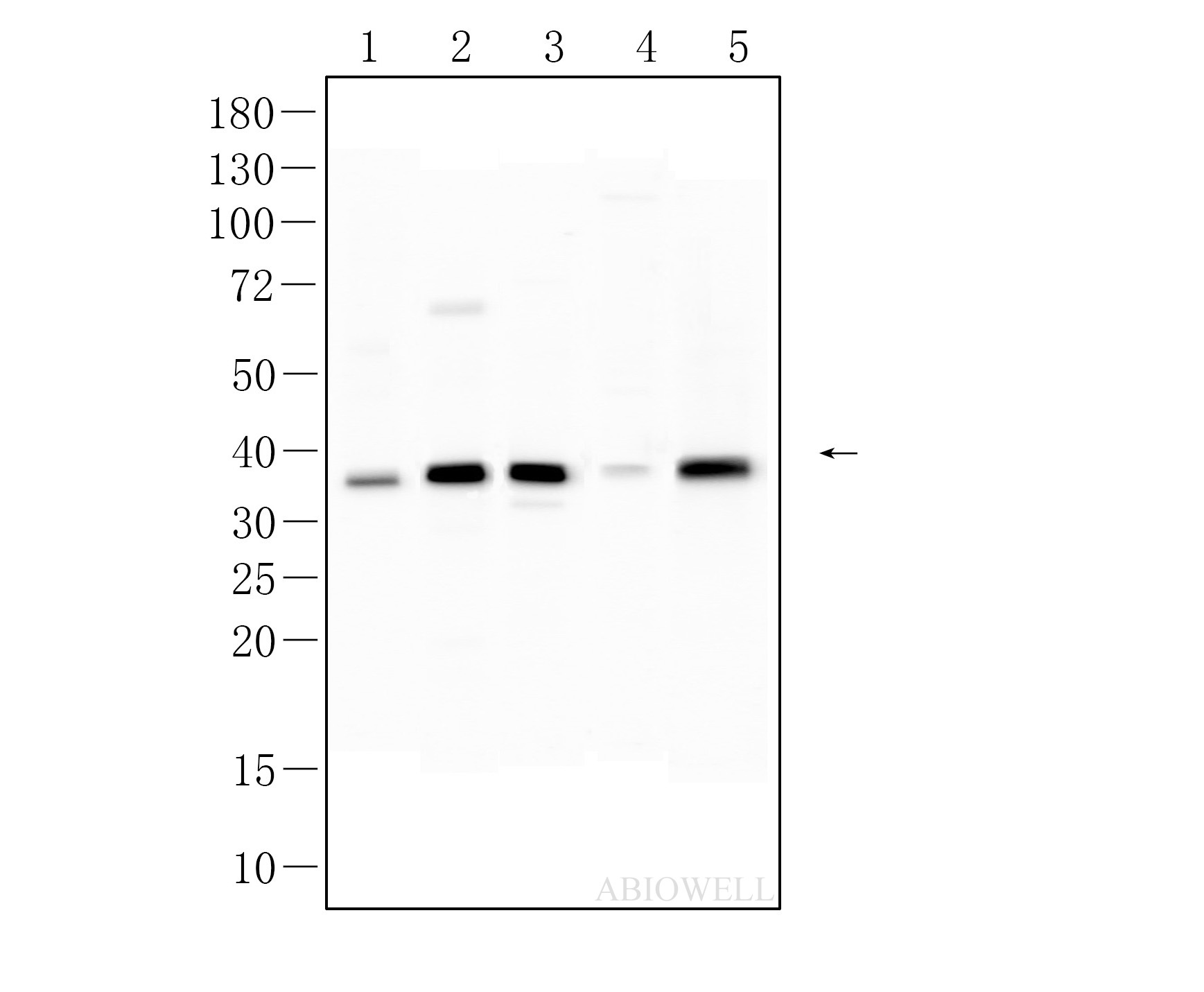
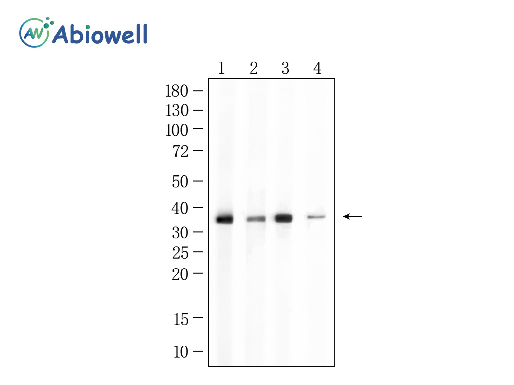
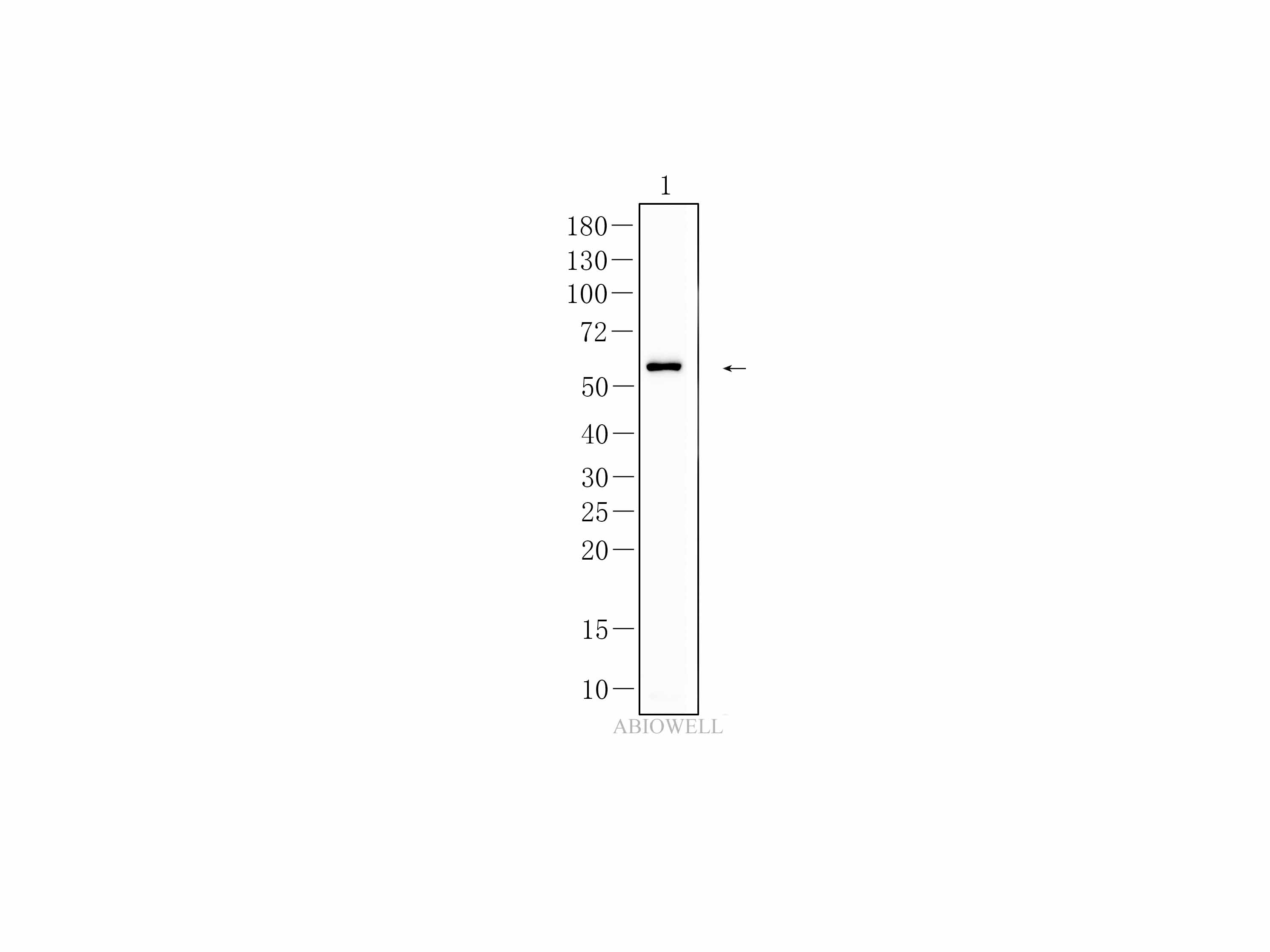
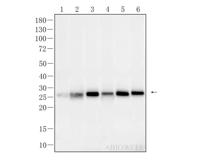
    xCT/SLC7A11 Recombinant Mouse Monoclonal Antibody [M43D07] - 2 Fig : Western blot analysis of xCT/SLC7A11 on different lysates. Proteins were transferred to a NC membrane and blocked with 5% NF-Milk in TBST for 1 hour at room temperature. The primary antibody (AWA00502, 1/1000) was used in TBST at room temperature for 2 hours. Goat Anti-Mouse IgG - HRP Secondary Antibody (AWS0002) at 1:5,000 dilution was used for 1 hour at room temperature.
    Positive control:
    Lane 1: Hela cell
    Lane 2: Mouse brain
    Predicted molecular weight:55 kDa
    Observed molecular weight:35-40 kDa,55 kDa
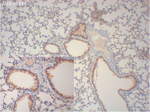
    xCT/SLC7A11 Recombinant Mouse Monoclonal Antibody [M43D07] - 3 Fig : Immunohistochemical analysis of paraffin-embedded Mouse-lung tissue with Mouse anti-xCT/SLC7A11 antibody (AWA00502) at 1/200 dilution.
    The section was pre-treated using heat mediated antigen retrieval with Sodium citrate buffer (pH 6.0) for 20 minutes. The tissues were blocked in 3% H2O2 for 15 minutes at room temperature, washed with ddH2O and PBS, and then probed with the primary antibody (AWA00502) at 1/200 dilution for 1 hour at room temperature. The detection was performed using an HRP conjugated compact polymer system(ABIOWELL, AWI0629). DAB was used as the chromogen. Tissues were counterstained with hematoxylin and mounted with DPX.
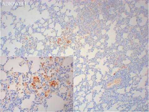
    xCT/SLC7A11 Recombinant Mouse Monoclonal Antibody [M43D07] - 4 Fig : Immunohistochemical analysis of paraffin-embedded Rat-lung tissue with Mouse anti-xCT/SLC7A11 antibody (AWA00502) at 1/200 dilution.
    The section was pre-treated using heat mediated antigen retrieval with  Sodium citrate buffer (pH 6.0) for 20 minutes. The tissues were blocked in 3% H2O2 for 15 minutes at room temperature, washed with ddH2O and PBS, and then probed with the primary antibody (AWA00502) at 1/200 dilution for 1 hour at room temperature. The detection was performed using an HRP conjugated compact polymer system(ABIOWELL, AWI0629). DAB was used as the chromogen. Tissues were counterstained with hematoxylin and mounted with DPX.

    引用文献 (4)

    FREE RADICAL BIOLOGY AND MEDICINE IF:8

    Mounting evidence highlights preeclampsia (PE) pathogenesis involves gut microbiota dysregulation and trophoblast ferroptosis. However, the mechanistic nexus bridging intestinal homeostasis and placental ferroptosis remains elusive. Here, we found that Faecalibacterium prausnitzii ( F. prausnitzii ) reduced blood pressure (SBP/DBP), proteinuria, and levels of anti-angiogenic (sFlt-1) and pro-inflammatory (IL-6, TNF-α) mediators in PE rats established using reduced uterine perfusion pressure (RUPP) surgery. Critically, F. prausnitzii elevated fetal survival rate, increased fetal length and weight, and restored placental length and weight. Moreover, F. prausnitzii restored intestinal barrier function (upregulated claudin-1, occludin, ZO-1/2). In both PE rats' placentas and hypoxia-induced HTR-8/SVneo cells, F. prausnitzii reduced ferroptosis markers (MDA, Fe 2+ , 8-OHdG, lipid ROS), while increasing GSH, GPX4, and SLC7A11 levels. Mechanistically, F. prausnitzii facilitated the transfer of intestinal-epithelial cell-derived extracellular vesicles (EVs) to the placenta and alleviated PE symptoms, by delivering NRP1 to inhibit trophoblast cell ferroptosis. Molecularly, NRP1 activates SLC7A11 both by directly binding to it and by stabilizing its mRNA via NSUN2-mediated m 5 C methylation. These findings demonstrate that F. prausnitzii alleviates PE by transporting EVs-encapsulated NRP1 through the gut-placenta axis, which in turn modulates the NSUN2/SLC7A11 pathway to suppress trophoblast ferroptosis. Our findings identify NRP1 as a core mediator of EV-dependent ferroptosis inhibition and highlight the therapeutic potential of targeting the gut microbiota or the NRP1/NSUN2/SLC7A11 axis for PE treatment.

    pubTime 2026-02-26
    Application
    CoIP,WB
    Specie
    Rat,Human
    Dilution
    1:1000
    MOLECULAR MEDICINE IF:6.4

    Background Radiation enteritis (RE) is a common complication in patients undergoing abdominal and pelvic radiotherapy. Despite the advancements in radiotherapy, effective treatments remain limited. WGX50, a bioactive compound from Sichuan pepper, has shown anti-inflammatory and antioxidant properties. This study investigates the protective effects of WGX50 on RE, focusing on its potential to reduce radiation-induced damage in the intestine. Methods Network pharmacology and molecular docking were used to identify the molecular targets of WGX50. In vitro, human intestinal epithelial cells (HIEC6) and colon cells (NCM460) were exposed to radiation and treated with WGX50. In vivo, C57BL/6 mice were administered WGX50 prior to radiation exposure. Various assays, including CCK-8, colony formation, flow cytometry, histopathology, and 16S rRNA sequencing, were performed to evaluate cell proliferation, apoptosis, oxidative stress, intestinal damage, and gut microbiota composition. Tissue transcriptome sequencing was conducted to explore differentially expressed genes. Results In vitro, WGX50 significantly mitigated radiation-induced cell damage, enhanced cell proliferation, and reduced apoptosis at non-toxic concentrations. In vivo, WGX50 treatment preserved intestinal morphology and reduced inflammatory infiltration in irradiated mice. WGX50 also protected goblet cells, maintaining mucin production and epithelial barrier function critical for intestinal homeostasis. Molecular docking, dynamics simulations and surface plasmon resonance (SPR) revealed stable binding of WGX50 to Epidermal Growth Factor Receptor (EGFR), key targets involved in oxidative stress regulation and ferroptosis inhibition. Mechanistically, WGX50 upregulated the EGFR-SLC7A11-GPX4 axis, suppressing ferroptosis and protecting intestinal cells. Additionally, 16S rRNA sequencing showed that WGX50 mitigated radiation-induced gut microbiota dysbiosis, preserving microbial diversity and promoting beneficial bacterial populations. Conclusion WGX50 demonstrates potent radioprotective effects by reducing oxidative stress, suppressing ferroptosis, and maintaining intestinal homeostasis, including goblet cell function and gut microbiota composition. These findings support WGX50’s potential as a novel therapeutic agent for the prevention and treatment of radiation enteritis.

    pubTime 2025-10-08
    Application
    IHC
    Specie
    Mouse
    Dilution
    1:200
    Frontiers in Chemistry IF:5.3

    A total of 22 terpenoids including one new triterpenoid ( 1 ), two new diterpenoids ( 2 and 3 ), and 19 known compounds ( 4 – 22 ) were isolated from the roots of Tripterygium regelii . Their structures were identified using NMR and HRESIMS techniques. Antiproliferative activity screening assays revealed that the new compound 2 , along with several known compounds, exhibits potential anti-colorectal cancer efficacy. Further investigations employing flow cytometry, immunofluorescence staining and electron microscopy demonstrated that compound 2 induces the generation of reactive oxygen species (ROS). This, in turn, promotes lipid peroxidation and mitochondrial damage, ultimately enhancing ferroptosis in colorectal cancer cells. These findings underscore the value of compound 2 as a potential candidate for anti-tumor drug development.

    pubTime 2026-02-20
    Application
    WB
    Specie
    Human
    Dilution
    BIOCHIMICA ET BIOPHYSICA ACTA-MOLECULAR BASIS OF DISEASE IF:4.2

    HIF-1 activation is protective in acute kidney injury (AKI), but its underlying mechanism is not fully understood. Stress-induced tRNA derived small RNAs play an emerging role in cellular processes. This study investigated the role of HIF-1 associated tiRNA-Lys-CTT-003 (tiR-Lys) in an AKI mouse model. Our sequencing results showed that ischemia can promote the production of renal tiR-Lys by activating HIF-1α. FG-4592, a HIF-1 inducer, can also upregulate the expression of tiR-Lys in renal tubular cells. Both overexpression of tiR-Lys and FG-4592 pre-treatment could improve mitochondrial damage and lipid peroxidation with alleviated renal function and morphological damage in cisplatin-induced AKI mice. While the anti-ferroptosis effect of FG-4592 were largely eliminated by tiR-Lys inhibitor. Notably, tiR-Lys directly alleviated cell death and MDA accumulation induced by the ferroptosis inducer Erastin, accompanied with restored expression of GPX4. RNA-Pulldown and RIP-qPCR results revealed that tiR-Lys can interact with the RNA-binding protein GRSF1.tiR-lys overexpression can preserve protein expression of GRSF1 decreased by cisplatin. Inhibiting Grsf1 via shRNA eliminated the upregulation of GPX4 by tiR-Lys. In conclusion, our study demonstrates that HIF-1α-induced tiR-Lys is protective in cisplatin-induced AKI, primarily by upregulating the level of GPX4 through interaction with GRSF1, thereby inhibiting ferroptosis in renal tubular epithelial cells.

    pubTime 2024-06-12
    Application
    WB
    Specie
    Mouse
    Dilution

    复制成功!

    内容已复制到剪贴板

    在线客服
    关闭
    小薇 薇薇 小艾 小奥
    联系销售
    技术支持

    image.png

    月琴艾碧维二维码.jpg