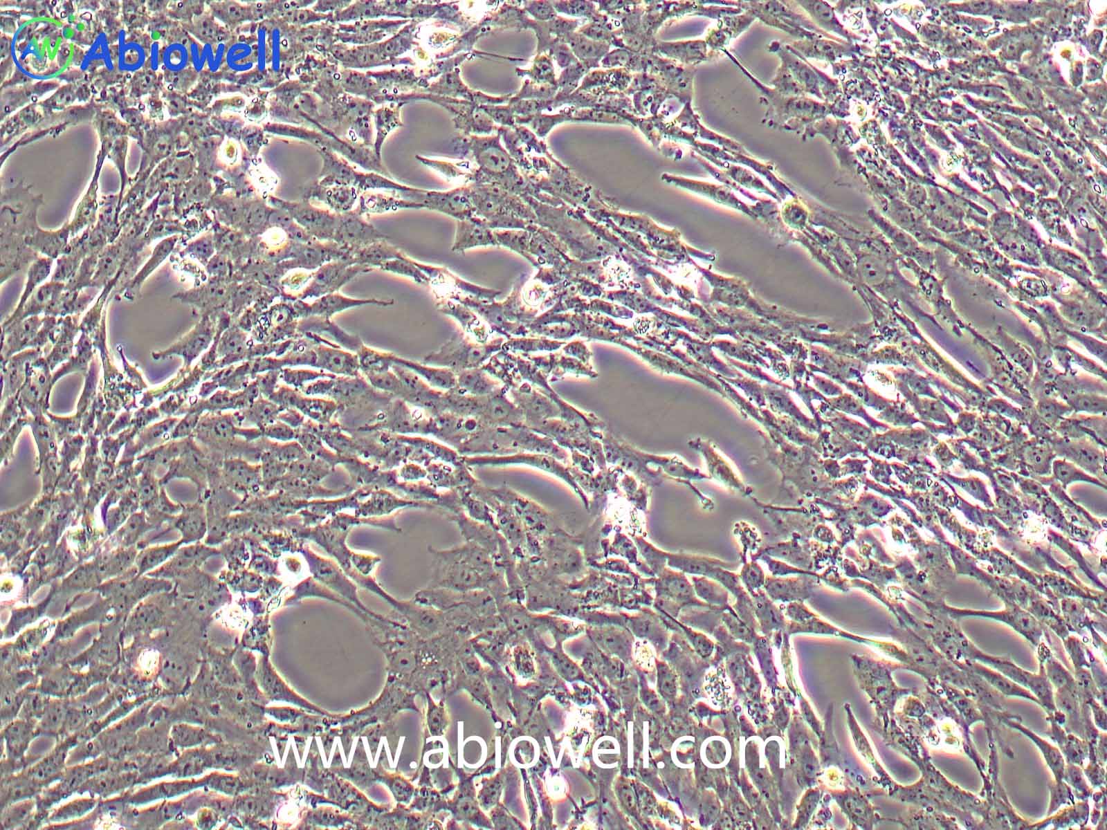
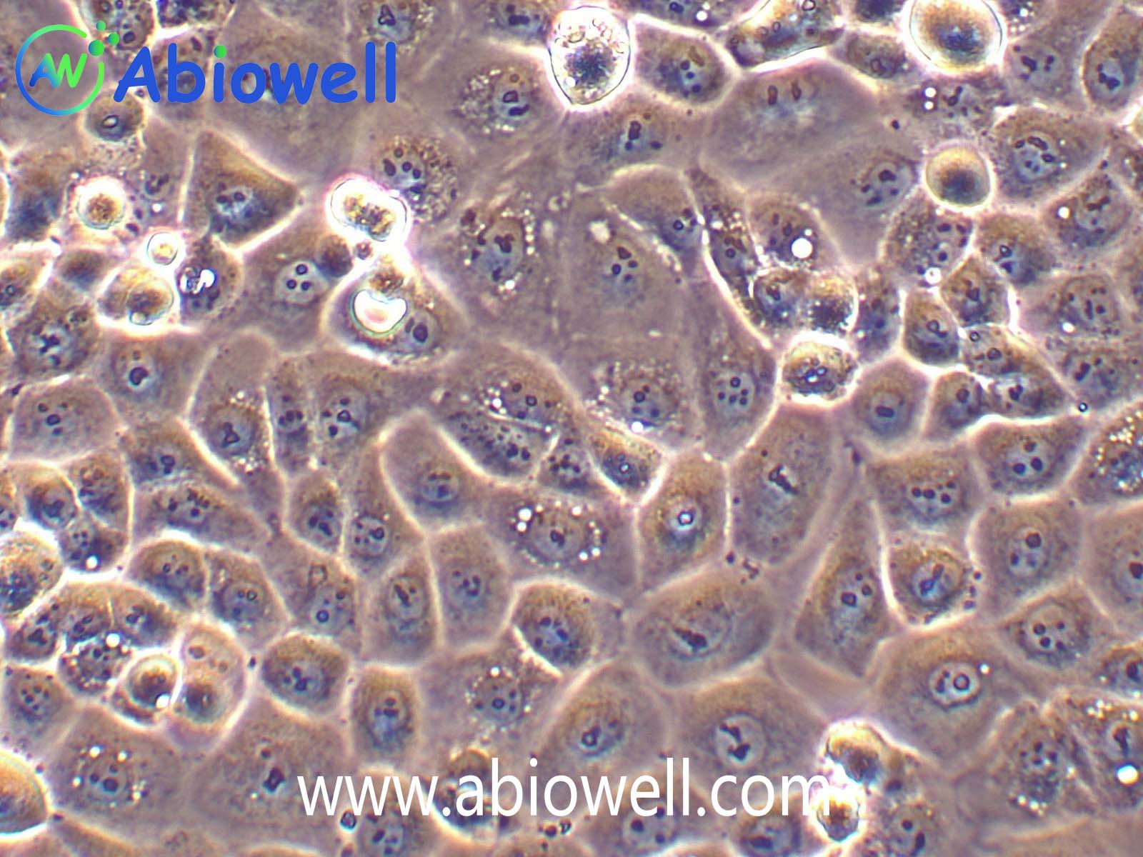
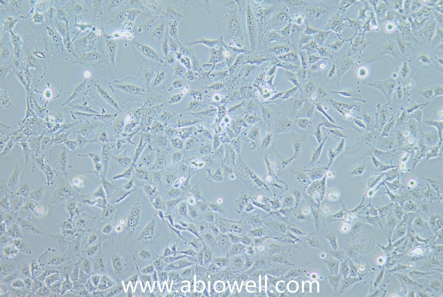
细胞及相关产品

联系我们CONTACT US

  • 172 6727 7467
    24小时服务热线
  • 销售邮箱

CAL-27 (人舌鳞癌细胞)(STR鉴定)

一键复制产品信息

货号:AW-CCH129

价格:¥1350

规格:1×10⁶cells

  • 产品概述
  • 细胞描述:

    该细胞系于1982年建立,从一位56岁白种男性的病变舌中间部分取得,该细胞具高密度颗粒状胞浆。

    细胞特性:

    1) 来源:舌

    2) 形态:上皮细胞样,贴壁生长

    3) 规格:1×10⁶cells

    4) 培养条件:DMEM+10%FBS+1%P/S (推荐货号AW-MC001)

                           空气,95%;二氧化碳,5%

                           37℃

    5)致瘤性: 可以,裸鼠皮下可成瘤。


    细胞接收后的处理:

    1) 收到细胞后,活细胞首先观察培养瓶是否完好,培养液是否漏液,培养基是否浑浊;冻存细胞是否干冰已挥发完,冻存管盖是否脱落,破碎,若有这类情况,请务必拍照记录,并于收货24h内与我们联系。

    2) 细胞处理:

    复苏的细胞:如果是T-25培养瓶活细胞,收到后请用75%的酒精对培养瓶表面进行消毒处理,然后转入培养箱中静置2~3h后再进行后续处理。

    备注:运输用的培养基不宜再次用来培养细胞,请按照说明书新配置完全培养基来培养细胞。  

    冻存细胞:如果是干冰运输的冻存细胞,收到后请立即转入液氮存储或者短暂(24h)放置-80度冰箱保存,或者直接进行细胞复苏。


    细胞复苏、传代及冻存流程参考:

    1、 细胞复苏

    1) 配制完全培养基:基础培养基+胎牛血清+双抗(特殊培养基特殊配置);

    2) 细胞复苏:取5ml完全培养基于15ml离心管中,37℃水浴锅预热,从液氮管(或者-80度冰箱)中快速取出冻存的细胞,放入37℃水浴锅中,摇晃使快速化冻(1min左右),然后将化冻的细胞和预热的培养基,移入超净工作台中,化冻的细胞加入到含预热培养基的15ml离心管中,1000rpm离心5min;

    3) 吸弃上清,得到细胞沉淀,用2ml完全培养基轻轻重悬细胞,加入到T25培养瓶中,做好标记,放入37℃,5%CO2饱和适度培养箱中培养(培养皿复苏效果更好);

    4) 24h后,观察细胞贴壁情况(未贴壁的即为死细胞--针对贴壁细胞),吸弃旧培养基,加入新鲜的预热(室温或37℃)的完全培养基,继续培养。

    2、 细胞传代

    1) 待细胞生长到80%-90%汇合度时,吸弃旧的培养基,加入1ml无菌PBS润洗一次,以去除残余的培养基及血清(血清含有胰酶的抑制因子),然后加入1ml 0.25%胰酶,37℃培养箱中消化(1~2min左右,不同细胞消化时间不同),取出细胞,镜下观察细胞至细胞皱缩变圆;

    2) 加入1ml完全培养基(含FBS)终止消化,轻轻拍打,使细胞脱落下来成单个细胞悬液,收集细胞于15ml无菌离心管中,1000rpm,离心5min;

    3) 收集细胞沉淀,完全培养基重悬,一分为二(可根据细胞生长速度调整比例),分别加入到2个新的培养瓶中,做好标记,放入培养箱中培养。

    3、细胞冻存

    1) 按照细胞传代方法,在超净工作台内消化收集细胞沉淀,取少量细胞用于计数;

    2) 用预冷的1ml冻存液(90%完全培养基+10%DMSO)或者无血清细胞冻存液重悬细胞,加入到1.2ml冻存管中,密度为1*106个/ml。

    3) 放入程序冻存盒,-80℃过夜后,转入液氮长期保存。

    参考文献 (3)

    CELLULAR SIGNALLING IF:3.7

    CNTNAP2 has two isoforms with confirmed expression: CNTNAP2–201 and CNTNAP2–203. While extensive research has focused on CNTNAP2–201 due to its association with various neurodevelopmental disorders, the role of CNTNAP2–203 in disease pathology remains largely unexplored. Oral squamous cell carcinoma (OSCC) is a major malignancy with high mortality, partially due to limited understanding of its molecular mechanisms. This study demonstrates that CNTNAP2–203 is predominantly expressed and significantly upregulated in OSCC tissues, and elevated CNTNAP2–203 is associated with poorer clinical outcomes. Functionally, CNTNAP2–203 promotes OSCC cell proliferation in vitro and tumor growth in vivo by positively modulating the activity of E2F transcription factor 1 (E2F1), a transcription factor that regulates G1/S cell cycle progression. Mechanistically, CNTNAP2–203 interacts with the epidermal growth factor receptor (EGFR), enhancing EGFR signaling and promoting OSCC tumorigenesis via the EGFR–E2F1 axis. Notably, OSCC cells with elevated CNTNAP2–203 exhibit increased sensitivity to Gefitinib (an EGFR tyrosine kinase inhibitor), either alone or in combination with Cisplatin, suggesting that patients with elevated CNTNAP2–203 may benefit from these treatments. In summary, this study not only elucidates the pathogenic role of CNTNAP2–203 in OSCC but also identifies it as a promising biomarker for guiding therapeutic strategies in OSCC management.

    Frontiers in Bioscience-Landmark IF:3.3

    Background: The importance of N6-methyladenosine (m6A) modification in tumorigenesis and progression have been highlighted. This study aimed to investigate the modification of insulin receptor substrate 1 (IRS1) by m6A and its role in oral squamous cell carcinoma (OSCC). Methods: Bioinformatics was employed to predict differential genes related to epithelial-mesenchymal transition (EMT) in OSCC. Seventeen pairs of OSCC and paracancerous tissue samples were collected. The impact of IRS1 on OSCC cell growth and EMT was evaluated. The fluctuations in IRS1 enrichment and the involvement of p53/Line-1 were investigated. Results: IRS1 was highly expressed in OSCC. IRS1 silencing decreased OSCC cell proliferation and increased apoptosis. IRS1 silencing hindered EMT by regulating related markers. IRS1 silencing upregulated p53 and downregulated Line-1 ORF1p. The p53 inhibition reversed the effects of IRS1 silencing and induced EMT in OSCC cells. Furthermore, the m6A modification of IRS1 was increased in OSCC cells. IRS1 were positively regulated by the m6A regulators methyltransferase-like 14 (METTL14) and YTH domain-containing protein 1 (YTHDC1). IRS1 bound to YTHDC1, and YTHDC1 knockdown inhibited the IRS1 nuclear export. The obesity-associated protein (FTO) negatively regulated IRS1, and FTO overexpression reversed the IRS1-induced OSCC tumor growth. Conclusions: m6A methylation-mediated IRS1 regulated EMT in OSCC through p53/Line-1. These findings provide potential therapeutic strategies for managing OSCC.

    MOLECULAR BIOTECHNOLOGY IF:2.4

    Oral squamous cell carcinoma (OSCC) is a common and highly lethal epithelial cancer. This study aimed to confirm the role of METTL3 in promoting OSCC and investigate its specific underlying mechanisms. Expression of the METTL3, YTH domain-containing family 2 (YTHDF2), and WEE1 were examined in normal oral epithelial cells and OSCC cells. Cell functions were examined after overexpressing WEE1 in OSCC cells. MeRIP-qPCR analysis was used to detect WEE1 m6A levels in HOK, SCC25, and CAL27 cells. WEE1 and its m6A levels were evaluated in OSCC cells by knocking down METTL3/YTHDF2, assessing the interaction between METTL3/YTHDF2 and WEE1. The impact of METTL3 and YTHDF2 downregulation on WEE1 mRNA stability was also investigated. The tumor weight and volume in a nude mouse model of OSCC after overexpression of WEE1 and YTHDF2 were measured. Expression of Ki-67 and WEE1 in OSCC tissue was detected using immunohistochemistry. Compared to normal oral epithelial cells, METTL3 and YTHDF2 were upregulated in OSCC cells, while WEE1 was downregulated, and there was a negative correlation between WEE1 and METTL3/YTHDF2 expression. WEE1 overexpression inhibited proliferation, invasion, and migration while promoting apoptosis in OSCC cells. METTL3 and YTHDF2 bound to WEE1 mRNA. METTL3/YTHDF2 knockdown increased WEE1 levels and WEE1 mRNA stability. METTL3 inhibition reduced WEE1 m6A levels. Inhibition of METTL3 weakened the interaction between YTHDF2 and WEE1 mRNA. In vivo, overexpression of WEE1 suppressed OSCC development, which was reversed by overexpression of YTHDF2. METTL3 facilitates the progression of OSCC through m6A-YTHDF2-dependent downregulation of WEE1. Graphic Abstract METTL3 promotes OSCC progression by enhancing WEE1 mRNA degradation in a m6A-YTHDF2-dependent manner. METTL3 enhances the binding of cytoplasmic YTHDF2 to m6A modification sites on WEE1 mRNA, thereby promoting WEE1 mRNA degradation and facilitating OSCC progression.

    复制成功!

    内容已复制到剪贴板

    在线客服
    关闭
    小薇 薇薇 小艾 小奥
    联系销售
    技术支持

    image.png

    月琴艾碧维二维码.jpg