辅助试剂

联系我们CONTACT US

  • 172 6727 7467
    24小时服务热线
  • 销售邮箱

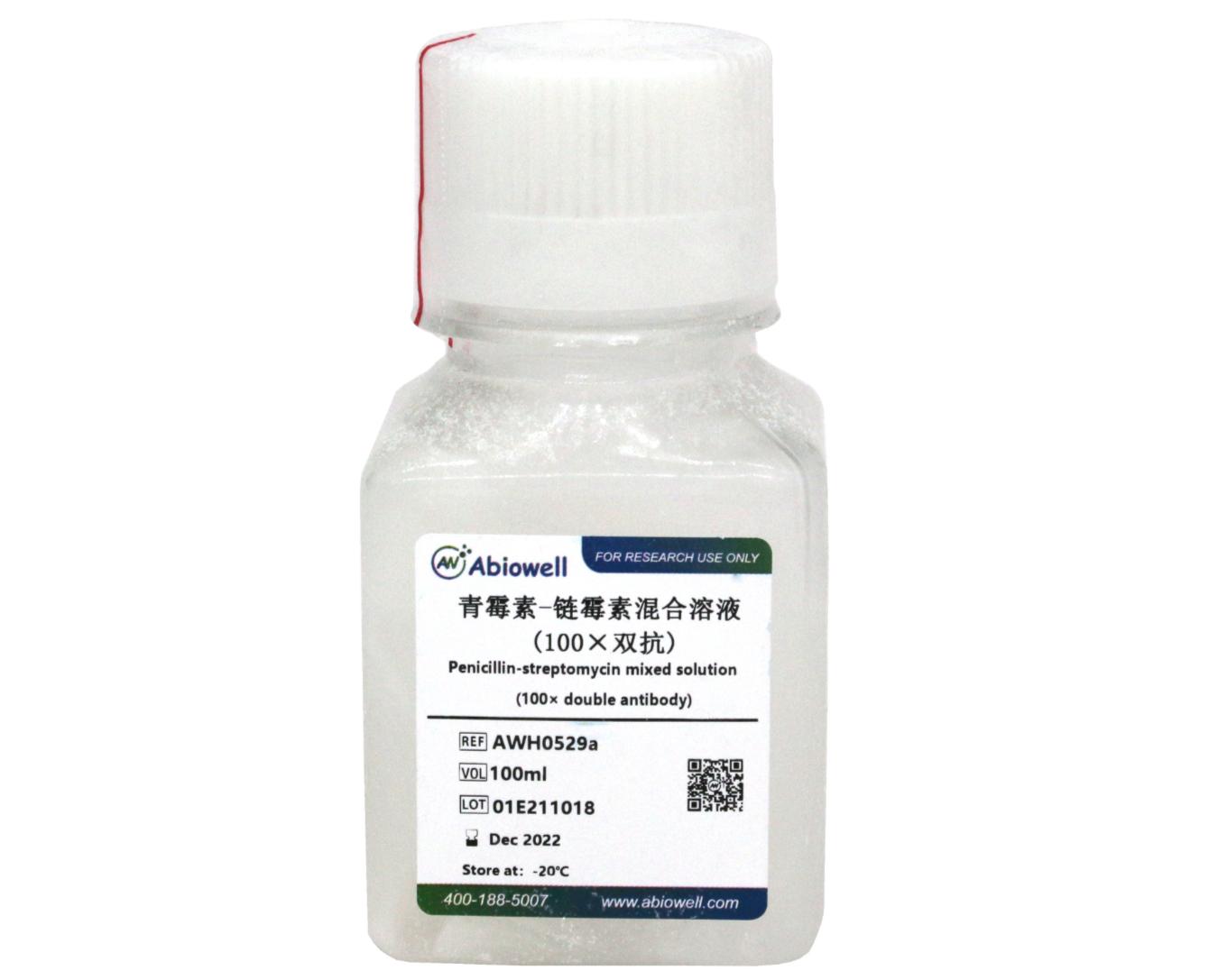
青霉素-链霉素混合溶液(100×双抗)

一键复制产品信息

货号:AWH0529

价格:¥80

规格:100ml

  • 产品概述
  • 青霉素-链霉素混合溶液(100×双抗)

    产品简介:

    青霉素-链霉素混合溶液(100×双抗)(Penicillin-Streptomycin Solution,100×)是专门用于细胞培养的双抗,经过滤除菌,可以直接添加到细胞培养液内。

    青霉素-链霉素混合溶液(100×双抗)中,青霉素的含量为10ku/ml,链霉菌的含量为10mg/ml。该溶液用0.9%NaCl或PBS配制。在细胞培养液中推荐的青霉素的工作浓度为100U/ml,链霉素的工作浓度为0.1mg/ml,即按照100倍稀释使用即可。一个包装即100ml青霉素-链霉素溶液(100×双抗)可以配制10L细胞培养液。

    操作步骤(仅供参考):

    青霉素-链霉素混合溶液(100×双抗)可以参考如下两种方法之一使用:

    1、 在无菌的细胞培养液中直接添加青霉素-链霉素溶液(100×双抗):按照每500ml细胞培养液添加5ml的比例加入青霉素-链霉素溶液(100×双抗),混匀即可使用。

    2、 配制细胞培养液时加入青霉素-链霉素溶液(100×双抗),然后再过滤除菌:配制细胞培养液时按照每配制1L细胞培养液加入10ml的比例加入青霉素-链霉素溶液(100×双抗),配制完成后过滤除菌即可使用。

    注意事项:

    1、 尽量减少反复冻融的次数,以免效率下降。

    2、 注意无菌操作,尽量避免污染。

    3、 为了您的安全和健康,请穿好实验服并佩戴一次性手套和口罩操作。

    4、 本产品仅限于专业人员的科学研究用,不得用于临床诊断或治疗,不得用于食品或药品,不得存放于普通住宅内。
    产品组成
    名称货号规格storage
    青霉素-链霉素混合溶液(100×双抗)AWH0529-100ml100ml-20℃

    注意:

    1.本产品仅供科研使用。请勿用于医药、临床诊断或治疗。食品及化妆品等用途。请勿存放于普通住宅区。

    2.为了您的安全和健康,请穿好实验服并佩戴一次性手套和口罩操作。

    3.实验结果可由多种因素影响,相关处理只限于产品本身,不涉及其他赔偿。


    参考文献 (54)

    Cell Metabolism IF:27.7

    Oligosaccharides are conventionally recognized as “passersby” in the small intestine. However, our research has reframed this understanding by uncovering a new function of oligosaccharide stachyose, which binds hydrophobic residues of membranous HSP90β on small intestinal epithelial cells, thus reprograming the exosomal miRNA profile. CRISPR-Cas9-mediated HSP90β knockout abolished the accumulation of stachyose on cell membrane and its regulatory effects on these miRNAs. Notably, stachyose’s regulation on these miRNAs is independent of its prebiotic role, as evidenced by the observation of stachyose-altered fecal miRNAs in pseudo-germ-free mice. These stachyose-altered miRNAs further shaped colonic microbiome, especially harboring Lactobacillus in mice. Thereinto, miR-30a-5p that was downregulated (Log 2 FC < −2) in both mice and human feces following stachyose treatment could specifically suppress the growth of Lactobacillus reuteri . These findings build a new regulatory axis of stachyose-intestinal miRNAs-gut microbiota and unveil a previously unknown mechanism underlying the direct “talk” of oligosaccharides to intestine epithelium via membranous HSP90β.

    Biomaterials Research IF:9.6

    Although tea consumption has been suggested to affect kidney stone formation, epidemiological evidence remains inconsistent, and the underlying molecular mechanisms are unclear. To assess the association between tea intake and kidney stone risk, we initially conducted a prospective cohort analysis of 481,393 participants from the UK Biobank and a 2-sample Mendelian randomization (MR) analysis. Our findings revealed that heavy tea drinkers (>5 cups/day) had a significantly reduced risk of kidney stones (hazard ratio: 0.79, 95% confidence interval [CI]: 0.72 to 0.86, P < 0.001), and MR analyses confirmed a causal association (inverse variance weighted OR: 0.45, 95% CI: 0.32 to 0.62, P < 0.001). We next explored the effect of epigallocatechin gallate (EGCG), the main bioactive component in tea, on calcium oxalate (CaOx) stone formation. EGCG was found to inhibit the glucose-regulated protein 94/phosphatidylinositol 3-kinase/protein kinase B (GRP94/PI3K/AKT) pathway in human proximal renal tubular epithelial cells, thereby attenuating CaOx crystal-induced oxidative stress and inflammation, and inhibiting crystal-cell adhesion. This finding aligned with the observation that the activated GRP94/PI3K/AKT pathway was positively associated with inflammation-related molecules in renal papillary tissues of CaOx stone formers. Moreover, to enhance renal targeting and therapeutic potential, we synthesized cell membrane-coated EGCG-loaded poly(lactic-co-glycolic acid) (TP-EGCG) nanoparticles, which enhanced renal EGCG delivery and substantially reduced CaOx crystal deposition in a mouse model of CaOx nephrolithiasis. In conclusion, tea consumption protects against kidney stone formation, an effect exerted by EGCG through the GRP94/PI3K/AKT axis, and our novel TP-EGCG nanoparticles show strong potential for targeted prevention and treatment.

    Cell Death & Disease IF:9

    Pathogenesis exploration and timely intervention of lung injury is quite necessary as it has harmed human health worldwide for years. Ficolin B (Fcn B) is a recognition molecule that can recognize a variety of ligands and play an important role in mediating the cell cycle, immune response, and tissue homeostasis in the lung. However, the role of Fcn B in bleomycin (BLM)-induced lung injury is obscure. This study aims to investigate the sources of Fcn B and its mechanism in BLM-induced lung injury. WT, Fcna -/- , and Fcnb -/- mice were selected to construct the BLM-induced lung injury model. Lung epithelial cells were utilized to construct the BLM-induced cell model. Exosomes that were secreted from alveolar macrophages (AMs) were applied for intervention by transporting Fcn B. Clinical data suggested M-ficolin (homologous of Fcn B) was raised in plasma of interstitial lung disease (ILD) patients. In the mouse model, macrophage-derived Fcn B aggravated BLM-induced lung injury and fibrosis. Fcn B further promoted the development of autophagy and ferroptosis. Remarkably, cell experiment results revealed that Fcn B transported by BLM-induced AMs exosomes accelerated autophagy and ferroptosis in lung epithelial cells through the activation of the cGAS-STING pathway. In contrast, the application of 3-Methyladenine (3-MA) reversed the promotion effect of Fcn B from BLM-induced AMs exosomes on lung epithelial cell damage by inhibiting autophagy-dependent ferroptosis. Meanwhile, in the BLM-induced mice model, the intervention of Fcn B secreted from BLM-induced AMs exosomes facilitated lung injury and fibrosis via ferroptosis. In summary, this study demonstrated that Fcn B transported by exosomes from AMs exacerbated BLM-induced lung injury by promoting lung epithelial cells ferroptosis through the cGAS-STING signaling pathway.

    Journal of Translational Medicine IF:7.4

    Background Right ventricle failure (RVF) is a progressive heart disease that has yet to be fully understood at the molecular level. Elevated M-type pyruvate kinase 2 (PKM2) tetramerization alleviates heart failure, but detailed molecular mechanisms remain unclear.ObjectiveWe observed changes in PKM2 tetramerization levels during the progression of right heart failure and in vitro cardiomyocyte hypertrophy and explored the causal relationship between altered PKM2 tetramerization and the imbalance of redox homeostasis in cardiomyocytes, as well as its underlying mechanisms. Ultimately, our goal was to propose rational intervention strategies for the treatment of RVF.MethodWe established RVF in Sprague Dawley (SD) rats by intraperitoneal injection of monocrotaline (MCT). The pulmonary artery pressure and right heart function of rats were assessed using transthoracic echocardiography combined with right heart catheterization. TEPP-46 was used both in vivo and in vitro to promote PKM2 tetramerization.Results We observed that oxidative stress and mitochondrial disorganization were associated with increased apoptosis in the right ventricular tissue of RVF rats. Quantitative proteomics revealed that PKM2 was upregulated during RVF and negatively correlated with the cardiac function. Facilitating PKM2 tetramerization promoted mitochondrial network formation and alleviated oxidative stress and apoptosis during cardiomyocyte hypertrophy. Moreover, enhancing PKM2 tetramer formation improved cardiac mitochondrial morphology, mitigated oxidative stress and alleviated heart failure.Conclusion Disruption of PKM2 tetramerization contributed to RVF by inducing mitochondrial fragmentation, accumulating ROS, and finally promoted the progression of cardiomyocyte apoptosis. Facilitating PKM2 tetramerization holds potential as a promising therapeutic approach for RVF.

    Translational Research IF:5.9

    Background Chronic obstructive pulmonary disease (COPD) is a chronic respiratory condition primarily caused by inhalation of harmful particles such as tobacco smoke. Cellular senescence serves as a key driver in its pathogenesis. Although endothelial progenitor cells (EPCs) have been shown to alleviate COPD by reducing inflammatory cell infiltration, the role and mechanisms underlying EPC senescence in this disease remain unclear. Methods A cigarette smoke (CS)-exposed COPD mouse model was established. Lung injury was assessed histologically, with concurrent quantification of neutrophil infiltration and cellular senescence levels in lung tissues. Pearson analysis evaluated the correlation between senescence severity and neutrophil numbers. In vivo neutrophil depletion was achieved using anti-Ly6G antibody, while GW4869 was used to inhibit exosome secretion from COPD-derived neutrophils. Neutrophils were then co-cultured with EPCs to assess their impact on EPC senescence and DNA damage. Proteomic analyses were employed to identify mechanisms of neutrophil-derived exosomes in COPD. Results COPD mice exhibited significant lung tissue damage, accelerated cellular senescence, and increased neutrophil infiltration. Senescence severity positively correlated with neutrophil proportion. Mechanistically, thrombospondin-1 (TSP-1) was highly expressed in COPD-derived neutrophils. Knockdown of neutrophil-derived exosomal TSP-1 alleviated EPC senescence. Furthermore, TSP-1 expression was regulated by transcription factor FOS, whereas TGF-β inhibition attenuated the promoting effects of TSP-1 overexpression on cellular senescence and lung injury in COPD mice. Conclusion This study demonstrates that neutrophil-derived exosomal TSP-1 aggravates EPC senescence and lung injury in COPD, revealing the pathogenic role of TSP-1 in disease progression and highlighting its potential as a therapeutic target.

    CELL BIOLOGY AND TOXICOLOGY IF:5.9

    Background Intervertebral disc (IVD) degeneration (IDD) represents a predominant origin of low back pain and disability, yet current therapeutic interventions remain suboptimal. Emerging evidence highlights autophagy activation as a therapeutic strategy against IDD. This study investigates the mechanistic interplay between N6-methyladenosine (m6A) modifications and autophagy dysregulation in IDD pathogenesis. Methods Bioinformatics analysis identified ring finger protein 41 ( RNF41 ) as a key autophagy-IDD intersection gene. Functional validation utilized tert-butyl hydroperoxide (TBHP)-treated human nucleus pulposus (NP) cells to assess RNF41’s effects on senescence (CDKN2A), autophagy (LC3-II/p62), apoptosis (TUNEL), inflammation (IL-18/IL-1β), and extracellular matrix (ECM) homeostasis (aggrecan/MMP). Key m6A regulators modulating autophagy were screened via correlation analysis. In vivo validation employed adeno-associated virus (AAV)-mediated methyltransferase-like 3 (METTL3)/RNF41 delivery in puncture-induced IDD rat models. Results RNF41 expression was downregulated in human IVD tissues. Overexpression of RNF41 mitigated TBHP-induced senescence, apoptosis, activated AMPK/mTOR-mediated autophagy, suppressed inflammation, and restored ECM balance. The autophagy inhibitor chloroquine (CQ) abolished the protective effects of RNF41 overexpression on degenerative NP cells. Mechanistically, METTL3/YTHDC1 co-regulation in degenerative NP cells mediated m6A hypermethylation of RNF41 mRNA, shortening its half-life via YTHDC1-dependent decay. Intradiscal METTL3-silencing AAV attenuated puncture-induced disc loss and histopathological degeneration, whereas RNF41-silencing AVV exacerbated ECM disruption and annular disorganization. Conclusion METTL3/YTHDC1-mediated m6A modification drives IDD progression by silencing RNF41 , thereby impairing autophagy and ECM integrity. Targeting this axis offers a clinically actionable strategy to delay disc degeneration, particularly in patients with early-stage IDD. This evidence establishes RNF41’s role as a theragnostic biomarker and therapeutic targe, enabling precision-guided interventional approaches.

    Frontiers in Immunology IF:5.7

    Objective: This study aimed to investigate the regulatory role of astrocyte-derived exosomes and their microRNAs (miRNAs) in modulating neuronal pyroptosis during cerebral ischemia.Methods: Astrocyte-derived exosomes were studied for treating cerebral ischemia in both in vitro and in vivo models. The effects of astrocyte-derived exosomes on neuroinflammation were investigated by analyzing exosome uptake, nerve damage, and pyroptosis protein expression. High throughput sequencing was used to identify astrocyte-derived exosomal miRNAs linked to pyroptosis, followed by validation via qRT‒PCR. The relationship between these miRNAs and NLRP3 was studied using a dual luciferase reporter assay. This study used miR-378a-5p overexpression and knockdown to manipulate OGD injury in nerve cells. The impact of astrocyte-derived exosomal miR-378a-5p on the regulation of cerebral ischemic neuroinflammation was assessed through analysis of nerve injury and pyroptosis protein expression.Results: Our findings demonstrated that astrocyte-derived exosomes were internalized by neurons both in vitro and in vivo. Additionally, Astrocyte-derived exosomes displayed a neuroprotective effect against OGD-induced neuronal injury and brain injury in the ischemic cortical region of middle cerebral artery occlusion (MCAO) rats while also reducing pyroptosis. Further investigations revealed the involvement of astrocyte-derived exosomal miR-378a-5p in regulating pyroptosis by inhibiting NLRP3. The overexpression of miR-378a-5p mitigated neuronal damage, whereas the knockdown of miR-378a-5p increased NLRP3 expression and exacerbated pyroptosis, thus reversing this neuroprotective effect.Conclusion: Astrocyte-derived exosomal miR-378a-5p has a neuroprotective effect on cerebral ischemia by suppressing neuroinflammation associated with NLRP3-mediated pyroptosis.Further research is required to comprehensively elucidate the signaling pathways by which astrocyte-derived exosomal miR-378a-5p modulates neuronal pyroptosis.

    BIOCHEMICAL PHARMACOLOGY IF:5.6

    Pancreatic cancer is highly challenging, with most patients developing intrinsic or acquired resistance to first-line chemotherapy drug gemcitabine (GEM). Although Matrix Metalloproteinase 28 (MMP28) is upregulated in pancreatic cancer and predicts a poor prognosis, its role in GEM resistance and molecular mechanism remain unclear. Here, we aimed to investigate the role of MMP28 in GEM resistance and molecular mechanism. First, differentially expressed genes in pancreatic cancer were identified through bioinformatics and validated in clinical samples and cells. MMP28 was significantly overexpressed in pancreatic cancer tissues and Capan-1 and PANC-1 cells, correlating with poor prognosis. Then, MMP28 knockdown was performed in Capan-1 and PANC-1 cells, followed by GEM treatment. Furthermore, in vivo experiments evaluated GEM sensitivity after MMP28 knockdown. The results showed that MMP28 knockdown enhanced GEM sensitivity both in vitro , reducing cell proliferation and survival, and in vivo , where tumor growth was significantly suppressed. Additionally, glycolysis-related changes were assessed. We revealed that glycolysis was implicated as a key pathway in this process, with reduced glucose uptake and lactate production observed after MMP28 knockdown. Protein-protein interaction analysis identified Staphylococcal nuclease domain-containing protein 1 (SND1) as a key interactor, and SND1 expression was upregulated in pancreatic cancer tissues. Moreover, MMP28 interacted with SND1 to regulate SND1′s recruitment of HK2 mRNA to promote glycolysis. However, overexpression of SND1 reversed the effects of MMP28 knockdown, restoring glycolysis and GEM resistance. In conclusion, MMP28 promoted tumor growth and GEM resistance in pancreatic cancer by regulating glycolysis via interaction with SND1.

    Cancer Cell International IF:5.3

    Background Triple-negative breast cancer (TNBC) is characterized by high invasiveness and metastasis potential. Ubiquitin carboxy-terminal hydrolase L1 (UCHL1) is strongly associated with breast cancer progression, although the underlying mechanisms are largely unknown. Methods The gene expression profiles of TNBC samples were downloaded from the TCGA database, and ubiquitination enzymes related to immune regulation were screened. UCHL1 expression in the TNBC tissues and in adipose-derived mesenchymal stem cells (ADSCs) stimulated in vitro with pro-inflammatory cytokines were analyzed. Exosomes were isolated from these stimulated ADSCs and transfected with scrambled (si-NC) or UCHL1-specific (si-UCHL1) siRNA constructs. TNBC cells were treated with the ADSCs-derived exosomes (ADSCs-Exos) and then co-cultured with macrophages or T cells. Finally, the tumorigenic potential of the ADSCs-Exos was evaluated by injecting the exosomes into mice bearing TNBC xenografts. Results UCHL1 was highly expressed in TNBC tissues and the stimulated ADSCs. The exosomes derived from stimulated ADSCs increased the viability and migration capacity of TNBC cells in vitro, and significantly increased Ki-67 expression through UCHL1. Furthermore, ADSCs-Exos induced M2 polarization of THP-1 monocytes by upregulating CD206 and Arg-1, and downregulating TNF-α and iNOS, and also decreased the proportion of CD3 + CD8 + T cells. Mechanistically, UCHL1 regulated the STAT3 and PD-L1 signaling pathways through HDAC6. Exosomes derived from the control and cytokine-stimulated ADSCs also promoted tumor growth in vivo, and increased the expression of UCHL1, CD206, HDAC6, STAT3, and PD-L1. However, UCHL1 knockdown reversed the pro-tumorigenic effects of the ADSCs-derived exosomes in vivo and in vitro. Conclusion Pro-inflammatory factors (IFN-γ + TNF-α) stimulating ADSCs-Exos enhance immune evasion in triple-negative breast cancer by regulating the HDAC6/STAT3/PD-L1 pathway via UCHL1 transporter. Thus, UCHL1 inhibition may enhance the response of TNBC to immunotherapy. Graphical Abstract

    Cancer & Metabolism IF:5.3

    Background Intrahepatic cholangiocarcinoma (ICC) is the second most common primary hepatocellular cancer. This study investigated whether ETV4 , ALYREF , and PKM2 affect glycolytic metabolism and ferroptosis, thereby potentially influencing ICC. Methods Bioinformatic analysis was used to explore the expression levels and prognosis of ETV4 , ALYREF , and PKM2 in ICC and their regulatory relationships were confirmed using in vitro experiments. Glycolytic metabolism and ferroptosis were examined, and chromatin immunoprecipitation and RNA immunoprecipitation experiments were performed to verify whether the ETV4 , PKM2 , and ALYREF could bind. The effect of ETV4/ALYREF on ICC was further confirmed by in vivo experiments. Results ETV4 , ALYREF , and PKM2 were highly expressed in ICC. Overexpressed (oe)-ETV4 and oe-PKM2 promoted cell migration and increased glucose (GLU) utilization and lactate and intracellular adenosine triphosphate (ATP) production. Addition of the ferroptosis inducer Erastin to the above groups revealed that sh-ETV4 and sh-ALYREF increased lipid reactive oxygen species (ROS), malondialdehyde (MDA), and Fe 2+ levels, and oe-PKM2 reversed these effects in the sh-ETV4 and sh-ALYREF groups. Oe-ETV4 promoted the expression of PKM2 , whereas sh-ALYREF inhibited the same. ETV4 could bind to ALYREF and PKM2 promoter, and ALYREF could promote the stability of PKM2 in an m5C-dependent manner. In vivo, ETV4 promotes tumor growth and the expression of proteins related to glycolytic metabolism by regulating ALYREF . Conclusion ETV4 promotes ICC development and ferroptosis resistance by facilitating glycolytic metabolism, and regulating PKM2 transcription by directly binding to the PKM2 promoter. Additionally, it mediates m5C-dependent PKM2 stabilization by directly binding to ALYREF . This study identified a new potential therapeutic target for ICC.

    复制成功!

    内容已复制到剪贴板

    在线客服
    关闭
    小薇 薇薇 小艾 小奥
    联系销售
    技术支持

    image.png

    月琴艾碧维二维码.jpg