产品介绍
组织在制作过程中,由于化学试剂的作用封闭了抗原,又由于热的作用致使部分抗原的肽链发生扭曲,致使在免疫组化的染色过程中不能将其显示出来,为了解决上述的问题,利用化学试剂和热的作用将这些抗原重新暴露出来或修正过来的过程称为抗原修复。柠檬酸盐、EDTA或Tris等缓冲液在热的条件下可以使被福尔马林屏蔽的抗原重新暴露出来,同时又不会对抗原表位造成破坏,从而提高抗原的检出率,降低背景染色,提高诊断的准确率。
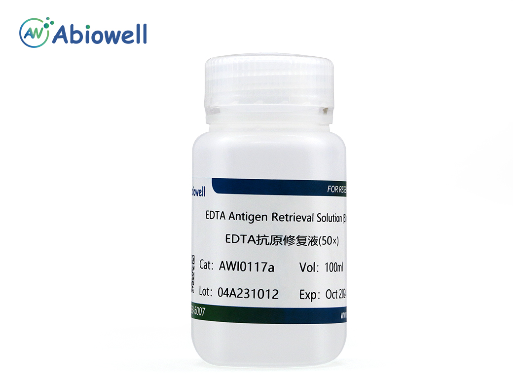
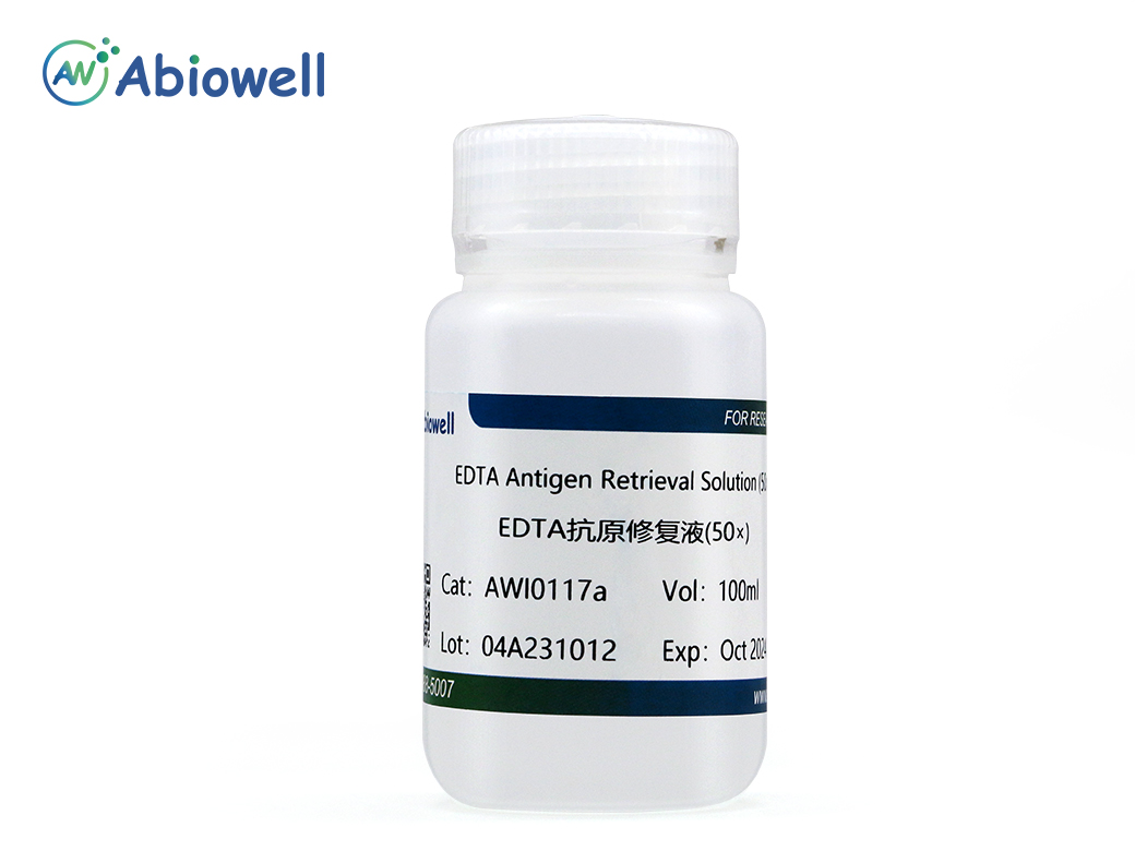
EDTA抗原修复液(EDTA Antigen Retrieval Solution, 50×)是一种常用的抗原修复液,可以有效去除醛类固定试剂导致的蛋白之间的交联,充分暴露石蜡切片等样品中的抗原表位,可以用于石蜡切片、冰冻切片等样品使用多聚甲醛、甲醛或其它醛类试剂固定后的抗原修复。细胞或组织用多聚甲醛、福尔马林或其它醛类试剂固定后,会导致蛋白之间的交联,遮蔽样品抗原位点,导致免疫染色信号减弱,甚至出现一些假阳性染色结果。抗原修复可以提高石蜡切片的免疫染色效果,亦可以不同程度的提高冰冻切片的染色效果。当冰冻切片免疫染色效果不理想时,考虑进行抗原修复。按照每个片子需要10ml抗原修复液(1×)计算,100ml抗原修复液(50×)可以用于500个样本的抗原修复。
产品规格
名称 | 货号 | 规格 |
EDTA抗原修复液(50×) | AWI0117a | 100ml |
EDTA抗原修复液(50×) | AWI0117b | 500ml |
保存条件
2-8℃
自备材料
1、 系列乙醇
2、 双蒸水或去离子水
3、 加热设备
4、 免疫染色洗涤液
使用方法
(一) 石蜡切片:
1、 脱蜡至水
①二甲苯3次,每次3~5min。
②无水乙醇脱水2次,每次3~5min。
③95%的乙醇,3~5min。
④90%的乙醇,3~5min。
⑤80%的乙醇,3~5min。
⑥70%的乙醇,3~5min。
⑦蒸馏水冲洗2次,每次3~5min。
2、 抗原修复
①用去离子水或双蒸馏水稀释EDTA Antigen Retrieval Solution(50×)至1×。
②将切片浸泡在抗原修复液(1×)中,95℃或沸水加热约10~30min。
③抗原修复液(1×)使用前需预热到95~100℃。如果使用微波炉加热,避免暴沸和过多的水分蒸发。随后大约在20~30min内冷却至室温。
3、 免疫染色洗涤液洗涤1~2次,每次3~5min。
4、 进行封闭等后续的免疫染色步骤。
(二) 冰冻切片:
1、 用去离子水或双蒸水稀释EDTA Antigen Retrieval Solution (50×)至1×。
2、 免疫染色洗涤液洗涤切片5min。
3、 将切片浸泡在抗原修复液(1×)中,95℃或沸水加热约10~30min。
4、 抗原修复液(1×)使用前需预热至95~100℃。如果使用微波炉加热,避免暴沸和过多的水分蒸发。随后大约在20~30min内冷却至室温。
5、 免疫染色洗涤液洗涤1~2次,每次3~5min。
6、 进行封闭等后续的免疫染色步骤。
(三) 其它样品:
其它样品参考石蜡切片或冰冻切片进行操作。
注意事项
1、 浸泡在抗原修复液(1×)中,最佳的加热时间需根据不同的样品和目的蛋白自行摸索。
2、 为了您的安全和健康,请穿好实验服并佩戴一次性手套和口罩操作。
3、 本产品仅限于专业人员的科学研究用,不得用于临床诊断或治疗,不得用于食品或药品,不得存放于普通住宅内。
参考文献 (2)
Background Hepatocellular carcinoma (HCC) ranks among the most aggressive malignancies worldwide, with poor outcomes attributed to delayed diagnosis and therapeutic limitations. Emerging evidence suggests that de novo lipogenesis (DNL) plays a crucial role in HCC progression and its interaction with the immune microenvironment. Methods We systematically analyzed DNL-related gene expression profiles from TCGA, GEO, ICGC-LIRI datasets, and our Xiangya HCC cohort ( n = 106) to construct a prognostic risk model. Through LASSO-Cox regression analysis, we identified six signature genes (G6PD, LCAT, SERPINE1, SOAT2, CYP2C9, and UGT1A10) that effectively stratified patients into distinct risk groups. We evaluated clinical characteristics, immune cell infiltration patterns, and differential therapeutic responses between high-risk and low-risk groups. Comprehensive validation included immunohistochemical analysis and Western blotting to assess expression levels of key model genes, along with multiplex immunofluorescence staining and single-cell RNA sequencing(scRNA-seq) to characterize immune microenvironmental differences between risk groups. Results We successfully established a robust six-gene prognostic signature (G6PD, LCAT, SERPINE1, SOAT2, CYP2C9, and UGT1A10) based on de novo lipogenesis pathways, which demonstrated excellent predictive performance (AUC: 0.78–0.82). The model revealed significant differences in immune infiltration patterns between risk groups, with the high-risk group exhibiting immunosuppressive characteristics characterized by increased Treg cell infiltration, while the low-risk group showed greater NK cell retention. Integrated scRNA-seq and our cohort validation further demonstrated that high-risk scores were associated with poorer response to immunotherapy but greater sensitivity to targeted therapies. These findings suggest that de novo lipogenesis-mediated immune evasion contributes to therapy resistance and worse prognosis in high-risk HCC patients, whereas low-risk HCC patients maintain an immunologically active microenvironment more amenable to immunotherapy. Conclusions This study provided a novel prognostic model for HCC, incorporating 6 representative DNLs. The model demonstrated the potential for predicting HCC prognosis and highlighted the involvement of immune cell infiltration and the association between risk scores and clinical therapy. Validation of model genes further supported the association between de novo lipogenesis and HCC development.
Background:Chronic heart failure (CHF) is a serious cardiovascular condition. Vascular peroxidase 1 (VPO1) is associated with various cardiovascular diseases, yet its role in CHF remains unclear. This research aims to explore the involvement of VPO1 in CHF.Methods:CHF was induced in rats using adriamycin, and the expression levels of VPO1 and cylindromatosis (CYLD) were assessed. In parallel, the effects of VPO1 on programmed necrosis in H9c2 cells were evaluated through cell viability assays, lactate dehydrogenase (LDH) level measurements, and analysis of receptor-interacting protein kinase 1/receptor-interacting protein kinase 3/mixed lineage kinase domain-like protein (RIPK1/RIPK3/MLKL) pathway-related proteins. The impact of CYLD on RIPK1 protein stability and ubiquitination was also investigated, along with the interaction between VPO1 and CYLD. Additionally, cardiac structure and function were assessed using echocardiography, Hematoxylin-eosin (HE) staining, Masson staining, and measurements of myocardial injury-related factors, including N-terminal prohormone of brain natriuretic peptide (NT-proBNP), Aspartate aminotransferase (AST), LDH, and creatine kinase-myocardial band (CK-MB).Results:VPO1 expression was upregulated in CHF rats and in H9c2 cells treated with adriamycin. In cellular experiments, VPO1 knockdown improved cell viability, inhibited necrosis and the expression of proteins associated with the RIPK1/RIPK3/MLKL pathway. Mechanistically, VPO1 promoted cardiomyocyte programmed necrosis by interacting with the deubiquitinating enzyme CYLD, which enhanced RIPK1 ubiquitination and degradation, leading to activation of the RIPK1/RIPK3/MLKL signaling pathway. At animal level, overexpression of CYLD counteracted the cardiac failure, cardiac hypertrophy, myocardial injury, myocardial fibrosis, and tissue necrosis caused by VPO1 knockdown.Conclusions:VPO1 exacerbates cardiomyocyte programmed necrosis in CHF rats by upregulating CYLD, which activates the RIPK1/RIPK3/MLKL signaling pathway. Thus, VPO1 may represent a potential therapeutic target for CHF.