辅助试剂

联系我们CONTACT US

  • 172 6727 7467
    24小时服务热线
  • 销售邮箱

BCA蛋白定量试剂盒

一键复制产品信息

货号:AWB0104

价格: ¥100

规格: 100T 250T 500T 2500T

  • 产品概述
  • BCA蛋白定量试剂盒

    产品简介:

    BCA蛋白定量法是目前常用的蛋白定量方法之一。本产品的原理是在碱性条件下,蛋白质将Cu2+还原为Cu+,Cu+与BCA试剂(Bicinchoninic Acid,二喹啉甲酸)形成紫色络合物,该络合物在562nm处有紫外吸收峰,与标准曲线对比,即可计算出待测蛋白的浓度。

    BCA法快速、灵敏、稳定。以BSA(牛血清蛋白)溶液为蛋白质标准品溶液,测定范围为20~2000μg/ml。

    注:

    ● 在低温条件或长期保存出现沉淀时,可搅拌或37℃温育使溶解,如发现细菌污染则应丢弃。

    ● 试剂A与试剂B用应密闭保存。

    操作步骤

    A.96孔酶标板测定:

    1、 BSA蛋白质标准工作液配制:用与待测蛋白样品一致的稀释液按下表稀释BSA标准品;

    2、 体积比为50∶1的比例配制适量BCA工作液,充分混匀;

    例子:BSA标准样品个数为7个,待测样品为2个,每个样品重复3次。

    96孔检测法所需BCA工作液总量为

    (7个BSA标准品样品+2个待测品样品)×3×0.2ml=5.4ml;

    再根据BCA-A和BCA-B体积比50∶1的比例计算所需量。但是由于加样过程中存在误差,建议多配2个孔的量。

    3、 96孔板检测过程:

    (1)将上述稀释好的BSA标准样品和待测样品各25μl分别加到做好标记的96孔板中,每个样品推荐做2-3个重复。

    (2)每个孔加入200μlBCA工作液,充分混匀,盖上96孔板盖,37℃孵育30min,冷却至室温,去除气泡,在3~5min内完成检测。

    (3)用酶标仪在540~590nm(建议是562nm,也可选择550/630nm)范围内检测96孔板中所有样品的吸光值。

    (4)根据上述BSA标准品浓度和吸光值绘制标准曲线,计算待测样品蛋白浓度。

    B.比色皿测定

    按照比色皿规格,与以上方法相同,适当按比例增加各溶液体积即可。

    使用注意事项:

    1、 由于该法检测时,吸光值会随时间延长而加深,因此,须在3~5min内完成检测,否则会影响蛋白浓度定量的准确性。

    2、 建议去除背景值后的吸光值读数绘制标准曲线。

    3、 使用温箱孵育时,应注意防止因水分蒸发导致的误差。

    4、 由于操作误差导致标准品读数严重偏离线性曲线的应该去除。

    5、 试剂A和试剂B混合后可能会出现浑浊,充分混匀后消失。

    6、 未知样品蛋白浓度可以从标准曲线方程计算得到,实际样品蛋白浓度需要乘以相应的稀释倍数。

    7、 如果得到的蛋白浓度不在检测范围内,请重新稀释样品后再次测定。

    8、 若样品中含有较高浓度的EDTA等螯合剂,建议选用Bradford法检测。

    注意事项: 

    1、 为了您的安全和健康,请穿好实验服并佩戴一次性手套和口罩操作。

    2、 本产品仅限于专业人员的科学研究用,不得用于临床诊断或治疗,不得用于食品或药品,不得存放于普通住宅内。
    产品组成
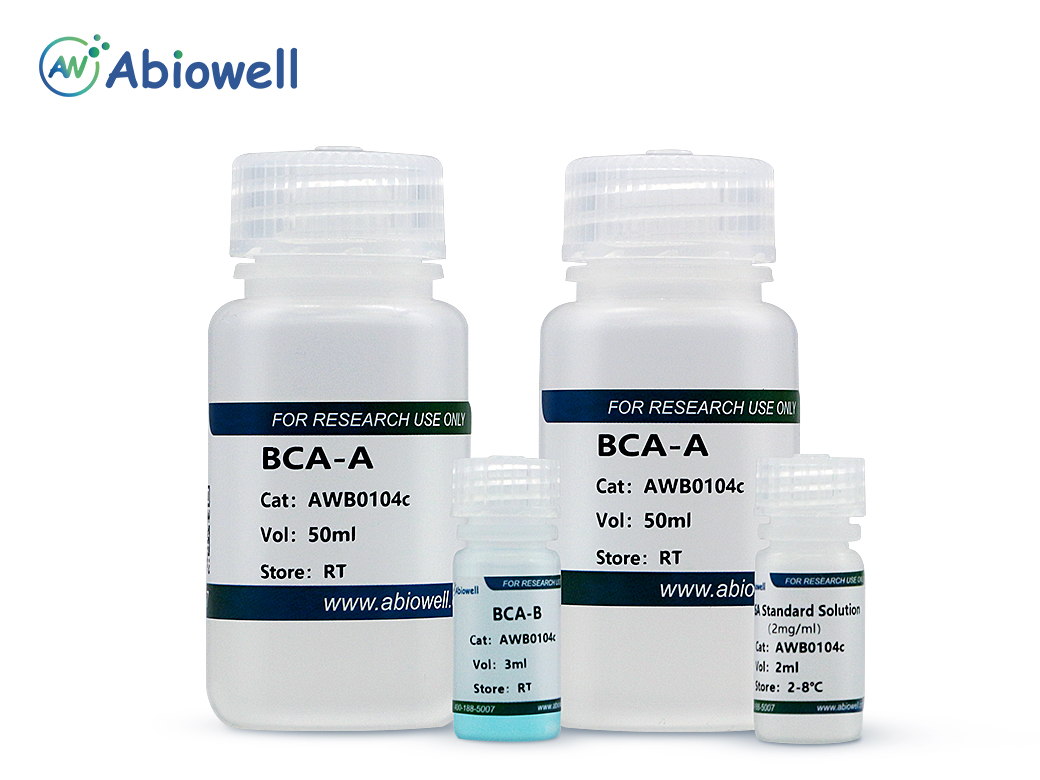
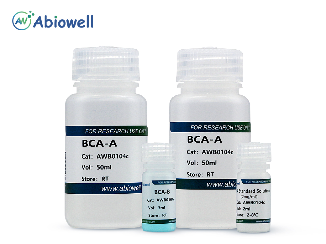
    名称AWB0104a(100T)AWB0104b(250T)AWB0104c(500T)AWB0104d(2500T)storage
    BCA-A20ml50ml100ml500mlRT
    BCA-B0.6ml1.5ml
    3ml15mlRT
    BSA Standard Solution0.5ml1ml2ml10ml2-8℃
    稀释BSA标准品
    编号稀释液用量(μl)BSA标准品用量(μl)BSA标准品最终浓度(μg/μl)
    A01002
    B2002001
    C200200(从B管取)0.5
    D200200(从C管取)0.25
    E200200(从D管取)0.125
    F200200(从E管取)0.0625
    G20000
    96孔检测法结果
    蛋白浓度(μg/ml)去除背景值后的吸光值原始吸光值
    000.0416(背景值)
    62.50.04490.08655
    1250.09180.1334
    2500.18960.2312
    5000.33310.3747
    10000.65240.694
    20001.16271.2043
    附表:(常见干扰物质的上限浓度)
    化合物浓度化合物浓度
    NP-401.0%葡萄糖10mM
    SDS1.0%蔗糖1M
    盐酸胍4MEDTA10mM
    Trition X-1001%β-巯基乙醇50μM
    甘氨酸1MDTT1mM
    乙酸盐0.2M尿素3M
    Tris0.1MDMSO5%
    NaCl1M甘油10%
    HCl/NaOH0.1MTES50mM
    HEPES0.1MMOPS50mM

    注意:

    1.本产品仅供科研使用。请勿用于医药、临床诊断或治疗。食品及化妆品等用途。请勿存放于普通住宅区。

    2.为了您的安全和健康,请穿好实验服并佩戴一次性手套和口罩操作。

    3.实验结果可由多种因素影响,相关处理只限于产品本身,不涉及其他赔偿。


    参考文献 (106)

    Advanced Science IF:14.1

    Secondary pneumonia, a common complication of sepsis-induced immunosuppression (SII), is closely linked to alveolar macrophage (AM) dysfunction primarily due to impaired glycolytic activity. However, the underlying molecular mechanisms remain unclear. In this study, it is found that exosomal RNA component of the mitochondrial RNA processing endoribonuclease (Rmrp), derived from type II alveolar epithelial cells (AEC-IIs), drives glycolytic defects and immune tolerance in AMs following cecal ligation and puncture (CLP) sepsis. Targeted depletion of Rmrp in either AEC-IIs or AMs alleviated SII and secondary pneumonia induced by Pseudomonas aeruginosa infection 48 h post CLP. Mechanistically, Rmrp interacts with and inhibits the ubiquitination and degradation of the RNA-binding protein zinc finger protein 36 (ZFP36). This results in ZFP36 upregulation, subsequently accelerating the decay of 6-phosphofructo-2-kinase/fructose-2,6-bisphosphatase 3 (Pfkfb3 ) mRNA by binding to its AU-rich elements in the 3′ untranslated region. The degradation of Pfkfb3 mRNA leads to impaired glycolysis and suppresses immune responses in AMs after sepsis. Additionally, it is found that exosomal Rmrp levels are correlated with AM immune tolerance and the prognosis of patients with sepsis. These findings highlight the critical role of AEC-II-derived exosomal Rmrp in the pathogenesis of SII and secondary pneumonia. Importantly, the study suggests that exosomal Rmrp may serve as a biomarker for predicting and managing SII in clinical settings.

    ACS Applied Materials & Interfaces IF:10.383

    In this work, a T2–T1 switchable superparamagnetic iron oxide nanoprobe with a pH/H2O2 dual response was obtained using a microemulsion method. This novel method for the controllable assembly of small iron clusters followed by their independent modification was reported, which could not be prepared by common synthetic methods. The size of the assembled nanoprobe was uniform and controllable, with a stable T2 magnetic resonance imaging (MRI) signal under a single condition. When the nanoprobe was exposed to the tumor environment, the higher H+ and H2O2 concentrations at the tumor site could dissociate the nanoprobe and redisperse into small iron clusters. When this occurred, the T2 MRI signal was converted into a T1 MRI signal, achieving specific detection of tumors by a pH/H2O2 dual-response T2–T1 MRI.

    Cell Death & Disease IF:9.6

    Ovarian cancer (OC) is often detected at an advanced stage and has a high recurrence rate after surgery or chemotherapy. Thus, it is essential to develop new strategies for OC treatment. This study tended to investigate the effects of endothelial cell-specific molecule 1 (ESM1) in OC. The impact of ESM1 on lipid metabolism was investigated through the regulation of ESM1 expression. Differential genes regulated by ESM1 were screened by mRNA sequencing. The role of autophagy in ESM1 regulation on lipid metabolism was explored using autophagy inhibitor chloroquine (CQ). Co-IP, dual-luciferase reporter assay, actinomycin D treatment assay, and others were used to analyze the mechanism of ESM1 regulation on lipid metabolism. The xenograft mouse model was constructed to explore the impact of ESM1 regulation on OC development. The regulatory mechanism of ESM1 in OC patient samples was verified by using microarray analysis and the Log-rank (Mantel-Cox) test. After ESM1 silencing, cholesterol synthesis decreased and lipolysis increased. mRNA sequencing revealed that ESM1 regulation on lipid metabolism was related to Beclin 1 (BECN1). In vitro experiments, ESM1 inhibited lipolysis by suppressing BECN1-mediated autophagy. BECN1 expression was regulated by the transcription factor Kruppel-like factor 10 (KLF10). The competitive binding between BECN1 and HSPA5 promoted the ubiquitination degradation of HMGCR, thereby inhibiting cholesterol production. The intervention experiment with exogenous cholesterol showed a positive correlation between m6A reader IGF2BP3 expression and cholesterol content. Mechanistically, IGF2BP3 regulated the stability of ESM1 mRNA. In vivo experiments, ESM1 modified by m6A methylation promoted cholesterol synthesis and inhibited lipolysis. High expression of ESM1 predicted poor prognosis in OC patients. ESM1 regulated lipid metabolism through IGF2BP3/ESM1/KLF10/BECN1 positive feedback, which was a promising target for OC treatment.

    Cell Death & Disease IF:9

    Pathogenesis exploration and timely intervention of lung injury is quite necessary as it has harmed human health worldwide for years. Ficolin B (Fcn B) is a recognition molecule that can recognize a variety of ligands and play an important role in mediating the cell cycle, immune response, and tissue homeostasis in the lung. However, the role of Fcn B in bleomycin (BLM)-induced lung injury is obscure. This study aims to investigate the sources of Fcn B and its mechanism in BLM-induced lung injury. WT, Fcna -/- , and Fcnb -/- mice were selected to construct the BLM-induced lung injury model. Lung epithelial cells were utilized to construct the BLM-induced cell model. Exosomes that were secreted from alveolar macrophages (AMs) were applied for intervention by transporting Fcn B. Clinical data suggested M-ficolin (homologous of Fcn B) was raised in plasma of interstitial lung disease (ILD) patients. In the mouse model, macrophage-derived Fcn B aggravated BLM-induced lung injury and fibrosis. Fcn B further promoted the development of autophagy and ferroptosis. Remarkably, cell experiment results revealed that Fcn B transported by BLM-induced AMs exosomes accelerated autophagy and ferroptosis in lung epithelial cells through the activation of the cGAS-STING pathway. In contrast, the application of 3-Methyladenine (3-MA) reversed the promotion effect of Fcn B from BLM-induced AMs exosomes on lung epithelial cell damage by inhibiting autophagy-dependent ferroptosis. Meanwhile, in the BLM-induced mice model, the intervention of Fcn B secreted from BLM-induced AMs exosomes facilitated lung injury and fibrosis via ferroptosis. In summary, this study demonstrated that Fcn B transported by exosomes from AMs exacerbated BLM-induced lung injury by promoting lung epithelial cells ferroptosis through the cGAS-STING signaling pathway.

    BIOMEDICINE & PHARMACOTHERAPY IF:7.5

    Background Cerebral ischemia-reperfusion injury (CIRI), a subsequent injury caused by thrombolytic reperfusion post ischemic stroke (IS). Naotaifang (NTF) formula, a novel traditional Chinese medicine (TCM) remedy against IS, was shown to exert beneficial effects in inhibiting inflammation and inhibiting lipid peroxide synthesis in our previous research. Purpose This study aimed to further explore the role of NTF in attenuating oxygen-glucose deprivation//reoxygenation (OGD/R)-induced inflammation and ferroptosis by regulating microglial M1/M2 polarization through the bone morphogenetic protein 6(BMP6)/SMADs signaling pathway. Methods BV2 microglia were used to establish an OGD/R model. The effects of NTF on inflammation and ferroptosis in OGD/R-injured BV2 cells were separately detected by immunofluorescence assay, fluorescent probe, DCFH-DA flow cytometry, enzyme-linked immunosorbent assay, and western-blot. Results The present results revealed that the M1 phenotype of microglia promoted the secretion of pro-inflammatory cytokines and aggravated ferroptosis and brain damage following OGD/R. However, an inhibitor of BMP6, LND-193189, reversed the aforementioned effects. Similarly, NTF promoted the shift of microglia from M1 to M2. Besides, NTF treatment effectively inhibited the expression of hepcidin, BMP6, SMADs and promoted the expression of ferroportin (FPN, SLC40A1) and γ‐L‐glutamyl‐L‐cysteinylglycine (glutathione or GSH) peroxidase 4 (GPX4). Conclusion Microglial M1/M2 polarization plays a pivotal role in inflammation and ferroptosis during OGD/R. The BMP6/SMADs signaling pathway is a potential therapeutical target of inflammation and ferroptosis induced by the transformation of microglia. Moreover, NTF could alleviate inflammation and ferroptosis through the BMP6/SMADs signaling pathway in OGD/R-injured microglia.

    BIOMEDICINE & PHARMACOTHERAPY IF:7.5

    Background Xin-tong-tai Granule (XTTG), a traditional Chinese medicine, has been used to treat atherosclerosis (AS), but its mechanism is poorly understood. Intriguingly, oxidative stress has been recognized as vital factors in the treatment of atherosclerosis. Purpose This study aims to explore the potential mechanism of XTTG for treating AS. Methods An in-vivo model of AS was established by feeding ApoE -/- mice with a high-fat diet (HFD), and the Human Aortic Vascular Smooth Muscle Cells (HAVSMCs) were induced by oxidized low-density lipoprotein (ox-LDL) in vitro. After treatment, the blood lipid levels and pathological aortic changes of each group were observed, and the cell proliferation and lipid droplet aggregation in each group were evaluated. The oxidative stress indicators such as malondialdehyde (MDA) and superoxide dismutase (SOD) levels and related NOX/ROS/NF-κB signaling pathway indicators were observed. Results XTTG improved blood lipid levels and pathological aortic changes of ApoE −/− mice with HFD feeding, inhibited HAVSMCs proliferation and lipid droplet aggregation induced by ox-LDL, reduced MDA content, increased SOD content, inhibited NOX4 and p22phox protein expression, downregulated ROS content, inhibited IKK-α, IKK-β, NF-κB protein and mRNA expression and the phosphorylation of NF-κB. Conclusion XTTG can inhibit NOX/ROS/NF-κB signaling pathway, reduce damages caused by oxidative stress, and exert anti-AS effects.

    Journal of Translational Medicine IF:7.5

    Background The molecular mechanisms underlying lymph node metastasis (LNM) in gastric cancer (GC) remain poorly understood. This study investigated HOXA9’s role in driving LNM via metabolic reprogramming. Methods Integrated analysis of gastric cancer RNA sequencing data and clinical specimens was performed. Functional validation involved HOXA9 overexpression and knockdown in AGS and HGC-27 cell lines, c-MYC silencing by siRNA, and glycolytic inhibition using 2-deoxyglucose (2-DG, 2.5 mM). In vitro assays evaluated proliferation (CCK-8), apoptosis (Annexin V/PI), migration/invasion (Transwell), lymphangiogenesis (HLEC tubulogenesis), and metabolism (Seahorse analyser). In vivo, effects were evaluated using a popliteal LNM mouse model ( n  = 6/group) and administered exogenous lactate (20 mM) to restore levels. Results HOXA9 was significantly upregulated in LNM-positive GC tissues (1.3-fold, p  = 0.0006) and predicted poor survival (HR = 1.57, p  = 1.7 × 10⁻⁵). HOXA9 overexpression enhanced GC cell proliferation (2.5-fold, p  < 0.0001), invasion (1.6-fold, p  = 0.0002), and migration (2.0-fold, p  < 0.0001), while suppressing apoptosis. Mechanistically, HOXA9 directly bound the c-MYC promoter, thereby upregulating glycolytic enzymes (HIF-1α, HK2, GLUT1, PDK1, LDHA) and increasing lactate secretion (1.7-fold, p  = 0.005). The resultant lactate-rich microenvironment stimulated lymphangiogenesis (1.4-fold, p  < 0.01) and endothelial cell migration (1.8-fold, p  < 0.001). These effects were significantly reversed by c-MYC knockdown or 2-DG treatment, with 2-DG reducing lymphangiogenesis by 37.56% ( p  < 0.0001). In vivo, HOXA9 knockdown reduced LNM burden (66% reduction in node volume, 83% lower metastasis rate), and this effect was markedly rescued by lactate supplementation. Conclusions HOXA9 promotes GC LNM by activating the c-MYC-glycolysis-lactate axis, which remodels the lymphatic niche. This axis represents a targetable pathway for GC therapy.

    Cell Death Discovery IF:7

    Pancreatic ductal adenocarcinoma (PDAC) is a highly lethal type of cancer with poor diagnosis and prognosis, and overcoming gemcitabine-resistant (Gem-R) is a major obstacle in its treatment. Given the important role of glutamine (Glu) metabolism in tumor drug resistance, we investigated the role and exact mechanism of transglutaminase type 2 (TGM2) in influencing PDAC sensitivity to gemcitabine. In this study, we found that TGM2 exhibited elevated expression levels in Gem-R cells and tissue samples from patients with clinically resistant PDAC. Mechanistically, downregulation of TGM2 suppressed the proliferation of Gem-R PDAC cells both in vitro and in vivo by modulating Glu metabolism. RNA sequencing analysis revealed that the mechanism by which targeting TGM2 inhibits drug resistance in Gem-R PDAC cells may be associated with purinergic receptor P2X7 (P2RX7) within the GO:0014049 pathway (positive regulation of glutamate secretion). P2RX7 is highly expressed in Gem-R PDAC cells and tissue samples, and it participates in Glu metabolism and mitophagy in Gem-R PDAC cells. Furthermore, Glu has also been found to induce mitophagy. Lastly, TGM2 and P2RX7 form a positive feedback regulatory loop, jointly regulating Glu metabolism and mitophagy, thereby promoting drug resistance in Gem-R PDAC cells. These data suggest that the TGM2-P2RX7 loop promotes Gem-R in PDAC by improving Glu metabolism and mitophagy, highlighting its potential as a crucial therapeutic target for PDAC.

    Cell Death Discovery IF:7

    Intestinal ischemia-reperfusion (II/R) injury represents a life-threatening and complex pathophysiological process that remains challenging to treat clinically, and emerging evidence suggests that ferroptosis plays an essential role in its pathogenesis. This study aimed to investigate whether extracellular vesicles derived from bone marrow mesenchymal stem cells (BMSC-EVs) can mitigate II/R-induced ferroptosis in a murine model. Using a bioinformatics database, we initially identified genes with abnormal expression patterns in II/R injury. Then, we confirmed the association between II/R injury, ferroptosis, and the HMGB1/SREBF2 axis through in vivo and in vitro experiments. To determine the role of HMGB1 in hypoxia/reoxygenation (H/R)-induced ferroptosis in Caco-2 cells, we transfected cells with either sh-HMGB1 or control sh-NC constructs and developed an H/R model in vitro. Subsequently, we examined factors regulating HMGB1-mediated ferroptosis in Caco-2 cells and assessed the effect of BMSC-EVs on this process. To further explore the mechanism underlying the protective effects of BMSC-EVs in II/R injury, we screened for miRNAs with reduced expression during II/R and verified their involvement. Among these, miR-378a-3p was identified as a candidate for regulating ferroptosis. To confirm its functional role, we treated II/R mice with BMSC-EVs overexpressing miR-378a-3p and assessed the outcomes. Our findings revealed that HMGB1, which is a key regulatory factor of ferroptosis, was significantly upregulated during II/R injury, and its knockdown alleviated H/R-induced ferroptosis in Caco-2 cells. We also found that SREBF2 directly regulates HMGB1 expression to promote H/R-induced ferroptosis in vitro. Importantly, BMSC-EVs alleviated II/R injury by suppressing ferroptosis in Caco-2 cells, and mechanistically, miR-378a-3p, a miRNA derived from BMSC-EVs, inhibited II/R-induced ferroptosis by modulating the SREBF2/HMGB1 axis. In conclusion, BMSC-EVs may exert protective effects against II/R injury by delivering miR-378a-3p, which regulates the SREBF2/HMGB1 axis to suppress ferroptosis, providing important insights into the pathological mechanisms underlying II/R injury and potential therapeutic strategies for its management.

    Cell Reports IF:6.9

    Bevacizumab (Bev) resistance limits therapeutic efficacy in ovarian cancer (OC) patients. We identified ESM1 as a key gene in Bev-resistant OC. ESM1 secreted by OC-resistant cell lines activates the ITGB1/FAK axis to induce neovascularization and Bev resistance. Additionally, ESM1 overexpression promoted the growth and Bev resistance of OC, lung, intestinal, and hepatocellular carcinoma tumors. Then, we identified TRIM28 as an upstream regulator that stabilizes ESM1 by promoting SUMOylation, inhibiting its proteasomal degradation. In OC mice, TRIM28 overexpression promotes angiogenesis and Bev resistance via ESM1-mediated ITGB1/FAK activation. This work unveils a new molecular pathway underlying Bev resistance in OC and proposes TRIM28 and ESM1 as potential therapeutic targets.

    复制成功!

    内容已复制到剪贴板

    在线客服
    关闭
    小薇 薇薇 小艾 小奥
    联系销售
    技术支持

    image.png

    月琴艾碧维二维码.jpg