辅助试剂

联系我们CONTACT US

  • 172 6727 7467
    24小时服务热线
  • 销售邮箱

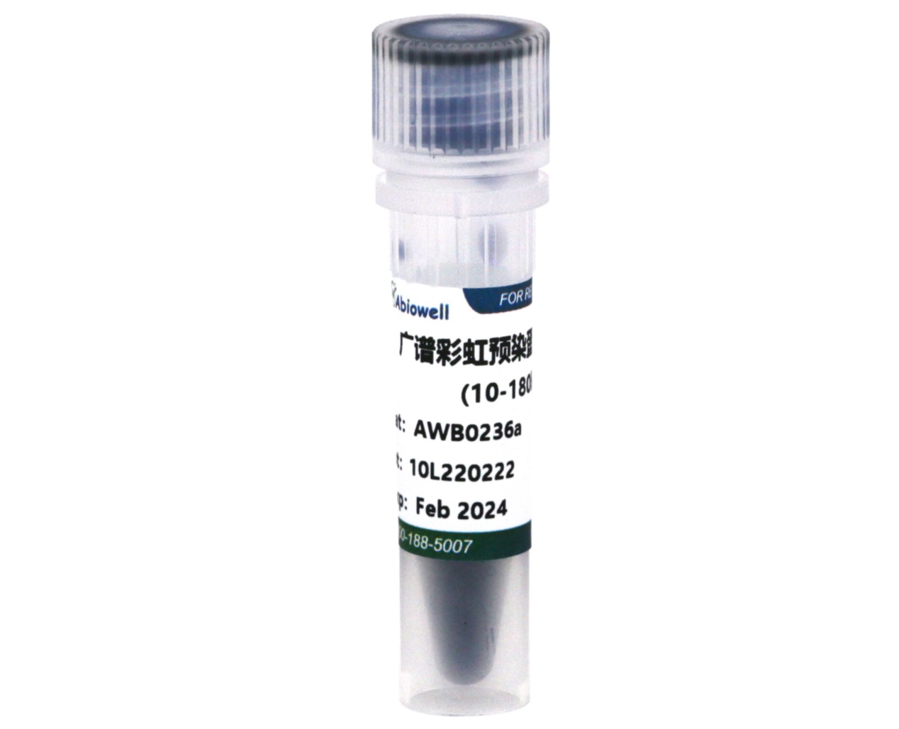
广谱彩虹预染蛋白Marker(10-180kD)

一键复制产品信息

货号:AWB0236

价格: ¥200 ¥350

规格: 250μL

  • 产品概述
  • 广谱彩虹预染蛋白Marker(10-180kD)

    产品介绍:

    本产品包内含11条预先染色的蛋白质条带,蛋白组分涵盖10kDa至180kDa。本产品经电泳(Tris-Glycine缓冲液)分离后,11条参考条带分别于蓝色、绿色或红色颜色基团共价键连接。为方便目测辨别分子量大小,绿色蛋白条带代表分子量25kDa;红色蛋白条带代表分子量72kDa。蛋白质常规分子量标记应用于辨别蛋白质电泳凝胶(SDS-PAGE)上的蛋白质大小及估算Western blot的转膜效率。

    本产品为即用型试剂,可直接使用,无需加热、稀释或添加还原剂。

    保存条件:

    4℃≤3个月,-20℃≤24个月

    使用方法:

    1、 加样前在室温数分钟解冻后轻轻摇匀,以确保溶液混合均匀;

    2、 取本产品5μl与实验样品同时进行聚丙烯酰胺凝胶电泳;建议有条件的实验室在初次使用本产品时可以根据自身的实验条件和实验习惯通过预实验确定合适的上样量,这样可以节约成本,同时获得效果更佳的实验图片。

    注意事项:

    1、 在低浓度胶中,低分子量蛋白会泳于染料前缘。

    2、 大分子量蛋白转印时建议延长转膜时间或加高转膜电压,如果效果不好建议微调转膜液配方,减少甲醇用量,添加少量SDS(终浓度不超过0.1%)。

    3、 预染蛋白质在不同的缓冲体系下有不同的表观分子量,如果在该缓冲体系中事先用非预染蛋白质标定,可以大致确定蛋白质分子量。

    4、 为了您的安全和健康,请穿好实验服并佩戴一次性手套和口罩操作。

    5、 本产品仅限于专业人员的科学研究用,不得用于临床诊断或治疗,不得用于食品或药品,不得存放于普通住宅内。

    实验结果:

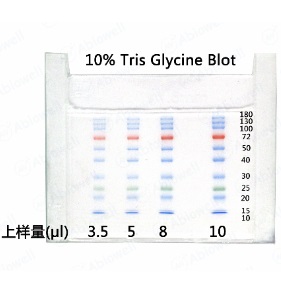
    Figure 1
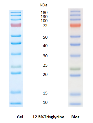
    AWB0236图片1.png

    Figure 2

    AWB0236图片2.png

    Marker常见案例及分析

    1. 蛋白Marker在凝胶上缺失部分条带?

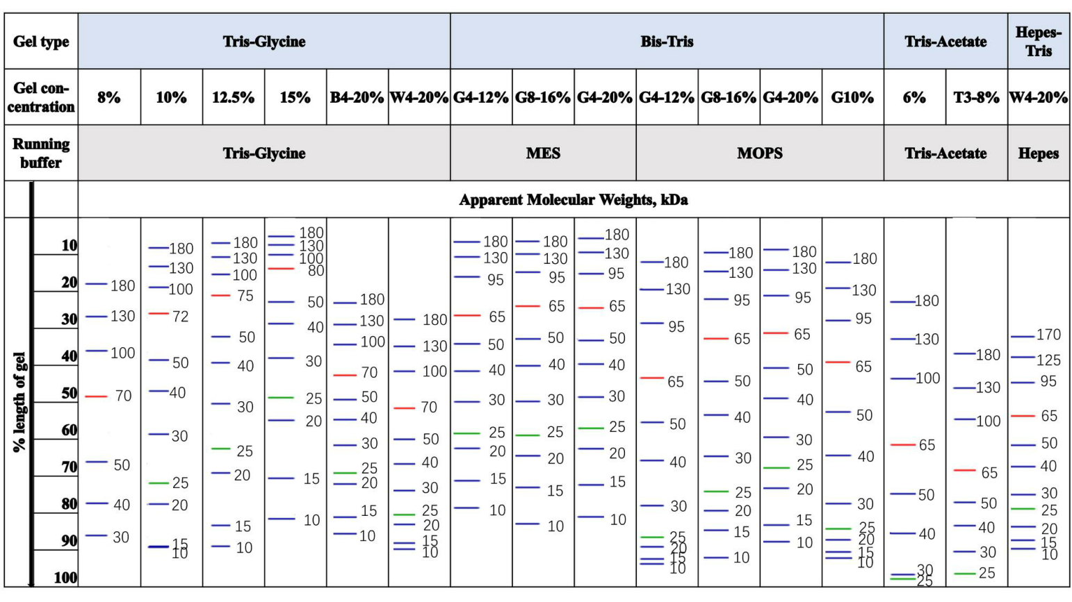
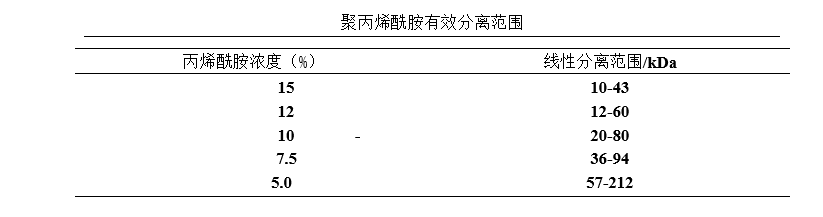
    检查使用的凝胶类型和百分比。根据蛋白分子量大小不同,使用不同孔径的聚丙烯酰胺凝胶。常用聚丙烯酰胺凝胶组分中双丙烯酰胺和丙烯酰胺的物质的量的比为1:29(或0.8:29.2),有效分离范围如下表。

    在Tris-Glycine胶中,通常12.5%的胶可以将10-180kDa的条带有效分开,10%的胶10/15/17kDa的条带会一起压缩在最前沿,8%的胶10/17/25会一起压缩在最前沿。
    AWB0236图片3.png

    2. 为什么你们的蛋白Marker跟其他品牌蛋白Marker条带指示大小不一致?换用不同凝胶,条带大小变化也不一样?

    SDS-PAGE中非预染蛋白marker受不同缓冲体系的影响小,而预染蛋白质marker由于多肽链上连接的染料基团受不同缓冲体系的影响比较大,因此预染蛋白marker在使用的不同缓冲体系中,需要用非预染蛋白marker进行标定。

    各个品牌条带指示大小不一致,可能有几个因素:(1)采用的标定方式和标示方法不一样。

    ①标定采用的非预染蛋白质marker不一样。分子量相同的蛋白由于氨基酸残基序列组成不同,与SDS的结合有差异,从而显示出不同的表观分子量。

    ②由于SDS-PAGE测量误差比较大,需要多次和多平行样测定。(2)各个品牌预染蛋白marker标记采用的染料不一样,导致在不同缓冲体系中的表观分子量变化不一样,有时差距极大。比如,某品牌蓝色条带在Tris-Glycine与Bis-Tris胶中比较,其表观分子量变化均超过10%以上,有的条带变化甚至超过20%以上。

    经测定,Abiowell预染蛋白质的蓝色和绿色条带表观分子量在不同的缓冲体胶中基本无变化(Tris-Glycine,Bis-Tris,Tris-acetate,Tricine)或变化极小(Hepes-Tris);橙色条带跟进口品牌变化基本一致,在Bis-Tris,Tris-acetate和部分改良TrisGlycine预制胶中表观分子量降低约10%。基本可以代替非预染marker。

    3. 在蛋白marker泳道中看到了一些额外的条带。可以提供一些建议吗?

    (1)上样时,请注意确保相邻样品泳道没有交叉污染。蛋白质上样过多,可能会飘散到别的泳道,导致产生额外的条带,这个问题在使用银染凝胶时尤为突出。

    (2)Marker储存不当或反复冻融会导致蛋白质降解,产生额外条带。Abiowell预染蛋白marker基本没有这种现象。

    4. 您们的蛋白marker的推荐上样体积是多少?

    0.75mm和1.0mm建议3-5ul,1.5mm建议8-10ul。

    5. 我可以将你们的预染蛋白marker用于非变性凝胶电泳吗?

    我们不建议将我们的预染蛋白质分子量标准品用于非变性凝胶电泳。因为它们已经变性(在 SDS 样品缓冲液中)和预还原(通过专有方法),预染marker其标示分子量不代表在非变性凝胶电泳中的样品分子量,但是可以作为一个电泳相对标尺。 

    6. 为什么预染蛋白marker电泳条带有时上面条带宽下面窄,不是一样的宽窄?

    请检查缓冲液的pH和更换pH标准品。   

    7. Abiowell的预染Marker产品稳定性如何?

    Abiowell产品非常稳定。通过我们的测试,Abiowell预染Marker25度放置35天几乎无变化;37度放置28天仅130kDa以上大分子稍变弱,不影响使用;50度放置9天仅130kDa以上大分子稍变弱,不影响使用;80度对Abiowell小分子影响小,大分子逐渐变弱。大分子逐渐变弱可能是高温溶解性不可逆变性,导致溶解性降低。

    8. 你们的预染蛋白marker是否不稳定,启用后条带逐渐消失降解,后只剩前沿的很深的蓝色条带?

    Abiowell产品非常稳定。产生这种现象的原因是由于实验操作不当或其他原因,在预染蛋白marker中引入了某种外源物质导致蛋白marker特异降解成小片段。解决方法:a注意实验操作习惯,使用干净的一次性的取样枪头,b多人使用时,蛋白marker启封后立即用干净的离心管和枪头分装。

    9. 使用蛋白marker,条带看起来弥散、模糊,怎么办?

    (1)配试剂尽可能使用超纯水。

    (2)选用合适浓度的凝胶并确认凝胶在正常有效期内。凝胶放置时间过长会影响分离效果,建议使用新配制的凝胶电泳。特别注意甲叉丙烯酰胺开封后多次称取会很快失效。

    (3)电压过高或电泳时间过长,产热过多导致电泳缓冲液温度升高。建议参照产品使用说明书。

    (4)电泳缓冲液陈旧,pH值不在缓冲范围内。建议使用新鲜的电泳缓冲液,为了获得理想的结果建议在使用前预冷缓冲液。

    (5)存储或取用不当,引入外源污染,导致蛋白降解。建议使用干净的枪头和EP管分装marker,用干净的枪头、缓冲液和电泳设备进行实验。

    (6)蛋白marker存储太久并存储不当,或很多次取用。 Abiowell产品不存在这个问题。

    10. 为什么有时候带型不整齐,呈向上弯曲状(“微笑状”)或向下弯曲状(“皱眉状”)?

    (1)“微笑状”条带可能是由于凝胶横向温度不均匀引起的,凝胶边缘附近的间隔条起到了散热器的作用。可调整实验温度,如将电泳设备置于4°C环境中,向外槽加入更多的缓冲液以散热,降低电泳功率,避免过热,过热能够引起条带扭曲,甚至会导致玻璃板碎裂。

    (2)“皱眉状”条带可能是由于电的不连续导致的不均匀电场(如玻璃板底部有气泡等)或凝胶厚度不一致引起的。凝胶边缘不完全聚合也能导致“皱眉状”条带。因此,在电泳开始前需要确保凝胶中或凝胶底部没有气泡,并且凝胶已经完全聚合。

    (3)如果在凝胶两侧泳道,由于边缘效应、离子强度不均等原因,均会导致带型变宽或弯曲。建议点样时将蛋白marker放在泳道中间或者在边缘点两个泳道marker。

    (4)凝胶配制不匀,凝胶内存有气泡,或点样孔中有细碎的残胶。

    (5)配胶过程中,分离胶和浓缩胶的界面不平。

    (6)电泳槽存在封闭不严的情况,导致电压不稳,出现跑胶条带倾斜。

    (7)检查试剂特别是甲叉丙烯酰胺是否失效。

    转膜:

    1. 预染蛋白Marker电泳后清晰,但是转膜后颜色变淡?

    通常情况下,转膜后Abiowell预染marker条带会更鲜亮,如果颜色变淡,可能有几种情况:

    (1)转印不完全

    操作不当,比如,转膜没有加冰袋,温度过高;胶和滤纸没夹紧。

    转膜条件不适合,转膜时间过长可能透膜,转膜时间过短,蛋白可能残留在胶内。

    膜没有充分活化,甲醇浓度太高易造成蛋白穿膜,转移到滤纸上;未活化可能也可能导致膜吸附蛋白能力不强。

    膜孔径不适合,建议小分子蛋白使用0.22um膜。

    (2)转印正常,可能使用了特殊的转膜缓冲液,建议使用通用转膜缓冲液,或品质有保障的商用转膜缓冲液。另外转膜液均不需要加SDS,若实验必须使用,建议SDS浓度不要超过0.02–0.04%。

    2. 洗膜的过程中颜色变浅?

    可能使用了比较特殊的缓冲液或者洗涤强度过强。建议使用通用缓冲液,或品质有保障的商用缓冲液,适当洗涤。较小孔径的膜可以在封闭和洗涤过程中更好地保留蛋白质。

    3. 为什么转膜后NC/PVDF膜上蛋白标品的条带会丢失?

    按标准操作流程,2.6-400kD都可正常转膜成功。

    4. 蛋白质marker里小分子蛋白质条带穿膜。该如何解决这个问题?

    按照标准操作流程,通常都可得到良好结果。如果出现蛋白条带穿膜而过的情况,重点检查以下几个因素:

    1)转印条件,建议适当降低电压、电流或缩短转印时间。

    2)确保转膜缓冲液的甲醇浓度和时长合适,Abiowell低分子量预染蛋白marker不需要加甲醇。转膜前使用浓度为10–20%的甲醇平衡PVDF膜,时间一般不超过30秒。

    3)SDS浓度适当。Abiowell小分子量和大分子marker转膜时均不需要SDS,若实验必须使用,建议SDS浓度不要超过0.02–0.04%。过多的SDS会阻碍蛋白质与膜的结合。

    4) 检查膜的质量、孔径和靶标蛋白质的大小。如果您的目标蛋白质小于10kDa,最好使用0.2μm及以下孔径的膜。
    产品介绍:

    分子量

    10,15,20,25,30,40,50,72,100,130,180kDa

    分子量种类

    11

    范围

    10  to 180

    染料颜色

    3 colors:Blue,Red,Green


    产品组成:

    名称

    货号

    规格

    广谱彩虹预染蛋白Marker(10-180kD)

    AWB0236a

    250μl


    注意:

    1.本产品仅供科研使用。请勿用于医药、临床诊断或治疗。食品及化妆品等用途。请勿存放于普通住宅区。

    2.为了您的安全和健康,请穿好实验服并佩戴一次性手套和口罩操作。

    3.实验结果可由多种因素影响,相关处理只限于产品本身,不涉及其他赔偿。


    参考文献 (4)

    INTERNATIONAL IMMUNOPHARMACOLOGY IF:4.7

    Objective Chronic insomnia can easily lead to clinical distress or cause mental, social, physical, educational, occupational, or other functional impairments. Considering the role of circadian rhythm in insomnia, we focused on exploring the action of miR-29a in regulating the PER2 gene in improving chronic insomnia. Methods LPS induces the expression of miRNAs targeting PER2 in HMC3 and PRM cells, which was verified by RT-qPCR. Poly-lactic- co -glycolic acid (PLGA) nanoparticles (NP) were used to encapsulate short hairpin (sh)-miR-29a to construct sh-miR-29a-NP. Morris water environment method was employed to establish a sleep deprivation rat model to investigate the therapeutic effects of sh-miR-29a-NP. Cell viability and levels of cell polarization factors were evaluated using CCK8 and ELISA, respectively. The Morris water maze test was applied to assess the learning and memory capabilities of the rats. Immunohistochemistry, immunofluorescence, and western blot were applied to test the expression of glial cell polarization, neuronal cell activation, apoptosis, and Period2 (PER2) /nuclear factor kappa B (NF-κB) axis proteins. Results miR-29a was significantly upregulated in LPS-induced HMC3 and PRM cells, with the most significantly altered miRNAs/PER2 interaction. In LPS-induced HMC3 and PRM cells, sh-miR-29a promoted the PER2 and CD206 expressions, and inhibited the expression of ionized calcium-binding adaptor molecule 1 (IBA-1), NF-κB, and CD86, while this effect was blocked by small interfering-PER2. Further in vivo experiments confirmed that PER2 and CD206 expression was reduced, while NF-κB, CD86, and IBA-1 expression were up-regulated in the hippocampal tissue of CSD rats. However, this effect was reversed by treatment with sh-miR-29a-NP. Treatment with sh-miR-29a-NP in CSD rats shortened the escape latency and increased the number of crossings over the original platform, while inhibited the expression of NLR Family Pyrin Domain Containing 3, caspase-1, Gasdermin D (GSDMD), and TUNEL signal in the hippocampal tissue. Conclusion The regulation of PER2/NF-κB pathway by sh-miR-29a-NP promoted M2 polarization of microglial cells and inhibited neuronal cell pyroptosis, thereby improving cognitive dysfunction in chronic insomnia.

    RESPIRATORY RESEARCH IF:4.7

    Background Interferon regulatory factor-1 (IRF1) is a transcription factor that plays a significant role in various biological processes, including inflammatory injury, viral infection, cell death, and immune responses, and it has been extensively studied in the context of different lung diseases. However, the mechanism underlying its involvement in lung fibrosis remains largely unknown. Methods Wild type (WT) mice, IRF1 global-null mice ( Irf1 −/− ) were subjected to a bleomycin-induced lung fibrosis model to enable examination of the role of IRF1 in lung fibrosis. Proteomic analysis of lung tissue from WT and Irf1 −/− mice treated with saline or bleomycin was performed to explore the mechanism of IRF1 in regulating lung fibrosis. Results In the bleomycin-induced fibrosis mouse model, increased expression of IRF1 was observed. Irf1 knockout mice displayed decreased lung fibrosis relative to WT mice following treatment with bleomycin. The protein expression of fibronectin, as assessed by the Western blot analysis of lung tissues, was downregulated in Irf1 −/− mice. We observed a similar reduction in collagen content using hydroxyproline detection. Histologically, there was less collagen deposition in the lungs of Irf1 −/− mice compared with WT mice. Proteomics data revealed that IRF1 may be involved in lung fibrosis via the regulation of ferroptosis. We determined that paraoxonase 1(PON1), a poorly characterized protein in lung fibrosis, was upregulated in Irf1 −/− mice following exposure to bleomycin. In vitro experiments revealed that IRF1 could regulate the level of GSH and MDA through PON1. We also determined that PON1 levels were lower in the plasma of IPF patients compared with healthy controls. Conclusion Our data highlight the importance of IRF1 in the fibrotic process, and PON1 may be a potential mediator of IRF1 in the progression of lung fibrosis.

    ARCHIVES OF BIOCHEMISTRY AND BIOPHYSICS IF:3

    Objective Stem cell therapy is expected to become a new treatment for central nervous system damage associated with perinatal hypoxic-ischemic encephalopathy (HIE), but the specific effects are unknown. This study explores the effects of human umbilical cord blood (HUCB) cells-secreted exosomal (HUCB-ex) MFG-E8 in neonatal rats with hypoxic-ischemic brain damage (HIBD), aiming to gain a theoretical foundation for the cure of perinatal HIE. Methods HIBD model was constructed in the Sprague Dawley rats (7-day-old). Rats were then intervened with 1×10 6 HUCB cells, HUCB-ex, or HUCB-ex oe-MFG-E8 , and HUCB-ex si-MFG-E8 . Primary microglia from rats were induced with oxygen-glucose deprivation and re-oxygenation (OGD/R), then co-cultured with either HUCB or primary neuronal cells, and subjected to treatment with HUCB-ex, HUCB-ex oe-MFG-E8 , HUCB-ex si-MFG-E8 , or Stattic. The expression of polarization factors and secreted factors in the microglia was measured using RT-qPCR, immunofluorescence, and western blot. Neuronal cell damage was assessed using MTT assays and flow cytometry. Behavioral impairments and brain tissue damage in the rats were evaluated using assays including the geotaxis reflex, cliff avoidance response, grip strength test, hematoxylin-eosin staining, TTC staining, and immunofluorescence. Results Early intervention with HUCB cells in HIBD rats increased test scores, decreased brain tissue weight, infarct area, as well as the IL-6, TNF-α, and IL-1β levels, and increased MFG-E8 levels. HUCB cells also decreased the levels of CD11b/c + CD45hi cells in HIBD rat brain tissue, and increased the levels of CD206 + CD11b/c + cells. In vitro experiments confirmed high expression of MFG-E8 in HUCB-ex. HUCB-ex si-MFG-E8 inhibited M2 polarization and induced neuronal cell injury through the SOCS3/STAT3 pathway. HUCB-ex and HUCB cells have equivalent therapeutic effects in HIBD rats. The treatment effectiveness of HUCB-ex was improved after delivering HUCB-ex oe-MFG-E8 , while was blocked after delivering HUCB-ex si-MFG-E8 . Conclusions HUCB-ex oe-MFG-E8 promoted M2 polarization of microglial and inhibited neuronal cell apoptosis through the SOCS3/STAT3 pathway, to alleviate behavioral disorders and brain tissue damage in HIBD rats.

    CURRENT MOLECULAR MEDICINE IF:2.5

    Background: Neuropathic pain is chronic and affects the patient’s life. Studies have shown that IRF5 and CXCL13/CXCR5 are involved in neuropathic pain; however, their interactions are unknown. Objective: In this study, a rat neuropathic pain model was constructed by inducing chronic compression injury (CCI). IRF5 recombinant lentiviral vector and CXCL13 neutralizing antibody were administered to investigate their action mechanisms in neuropathic pain. Consequently, the new strategies for disease treatment could be evolved. Methods: The CCI rats were intrathecally injected with recombinant lentivirus plasmid LV-IRF5 (overexpression), LV-SH-IRF5 (silencing), and CXCL13 neutralizing antibody. Mechanical withdrawal threshold (MWT) and thermal withdrawal latency (TWL) were measured. The tumor necrosis factor (TNF)-alpha, interleukin (IL)-1β, and IL-6 levels were recorded via the enzyme-linked immunosorbent assay (ELISA). The spinal cord was stained using hematoxylin–eosin (HE). The binding of IRF5 to CXCL13 was analyzed by chromatin immunoprecipitation (ChIP) and dual luciferase reporter assay. The IRF5, neuronal nuclei (NeuN), CXCL13, and CXCR5 expressions were detected through quantitative real-time polymerase chain reaction and Western blot. Results: The MWT and TWL values in the CCI group were lower than in the Sham group. The expressions of CXCL13, CXCR5, and IRF5 in CCI rats were gradually increased with the modeling time. IRF5 silencing suppressed the expression of NeuN and lumbar enlargement in CCI rats and promoted MWT and TWL. Moreover, IRF5 silencing inhibited the expressions of CXCR5 and CXCL13 genes and down-regulated the expression levels of inflammatory factors. IRF5 was directly and specifically bound with the endogenous CXCL13 promoter and thus regulated it. IRF5 overexpression exacerbated the disease phenotype of CCI-induced neuropathic pain in rats. Administration of CXCL13 neutralizing antibodies reversed the IRF5 overexpression effects. Conclusion: The IRF5 silencing alleviated neuropathic pain in CCI rats by downregulating the pain threshold, inflammatory cytokine levels, and CXCL13/CXCR5 signaling. IRF5 overexpression exacerbated the disease parameters of CCI-induced neuropathic pain in rats; however, they were reversed by neutralizing antibodies against CXCL13.

    复制成功!

    内容已复制到剪贴板

    在线客服
    关闭
    小薇 薇薇 小艾 小奥
    联系销售
    技术支持

    image.png

    月琴艾碧维二维码.jpg