辅助试剂

联系我们CONTACT US

  • 172 6727 7467
    24小时服务热线
  • 销售邮箱
  • Fig: Fluorescence immunohistochemical analysis of Mouse-ileum tissue (Formalin/PFA-fixed paraffin-embedded sections). with Rabbit anti-Integrin alpha 2 antibody (AWA12676) at 1/200 dilution.

The immunostaining was performed with the TSA Immuno-staining Kit (ABIOWELL, AWI0688). The section was pre-treated using heat mediated antigen retrieval with EDTA buffer (pH 9.0) for 20 minutes. The tissues were blocked in 5% BSA for 60 minutes at 37℃, washed with ddH2O and PBS, and then probed with the primary antibody (AWA12676) at 1/200 dilution for 1 hour at room temperature. The detection was performed using an HRP conjugated compact polymer system followed by a separate fluorescent tyramide signal amplification system (green). DAPI (blue, AWC0291) was used as a nuclear counter stain. Image acquisition was performed with  Slide Scanner.
  • Fig: Fluorescence immunohistochemical analysis of Rat-Colon tissue (Formalin/PFA-fixed paraffin-embedded sections). with Rabbit anti-Integrin alpha 2 antibody (AWA12676) at 1/200 dilution.

The immunostaining was performed with the TSA Immuno-staining Kit (ABIOWELL, AWI0688). The section was pre-treated using heat mediated antigen retrieval with EDTA buffer (pH 9.0) for 20 minutes. The tissues were blocked in 5% BSA for 60 minutes at 37℃, washed with ddH2O and PBS, and then probed with the primary antibody (AWA12676) at 1/200 dilution for 1 hour at room temperature. The detection was performed using an HRP conjugated compact polymer system followed by a separate fluorescent tyramide signal amplification system (green). DAPI (blue, AWC0291) was used as a nuclear counter stain. Image acquisition was performed with  Slide Scanner.
  • Fig: Fluorescence immunohistochemical analysis of Rat-Liver  Tissue (Formalin/PFA-fixed paraffin-embedded sections). with Rabbit anti-STAT3(phospho-S727)  antibody (AWA12673) at 1/200 dilution.

The immunostaining was performed with the TSA Immuno-staining Kit (ABIOWELL, AWI0688). The section was pre-treated using heat mediated antigen retrieval with EDTA buffer (pH 9.0) for 20 minutes. The tissues were blocked in 5% BSA for 60 minutes at 37℃, washed with ddH2O and PBS, and then probed with the primary antibody (AWA12673) at 1/200 dilution for 1 hour at room temperature. The detection was performed using an HRP conjugated compact polymer system followed by a separate fluorescent tyramide signal amplification system (green). DAPI (blue, AWC0291) was used as a nuclear counter stain. Image acquisition was performed with  Slide Scanner.
  • Fig: Fluorescence immunohistochemical analysis of Mouse-bladder  tissue (Formalin/PFA-fixed paraffin-embedded sections). with Rabbit anti-PKR (phospho Thr446) antibody (AWA40532) at 1/200 dilution.

The immunostaining was performed with the TSA Immuno-staining Kit (ABIOWELL, AWI0688). The section was pre-treated using heat mediated antigen retrieval with EDTA buffer (pH 9.0) for 20 minutes. The tissues were blocked in 5% BSA for 60 minutes at 37℃, washed with ddH2O and PBS, and then probed with the primary antibody (AWA40532) at 1/200 dilution for 1 hour at room temperature. The detection was performed using an HRP conjugated compact polymer system followed by a separate fluorescent tyramide signal amplification system (green). DAPI (blue, AWC0291) was used as a nuclear counter stain. Image acquisition was performed with  Slide Scanner.
  • Fig: Fluorescence immunohistochemical analysis of Rat-lung (Formalin/PFA-fixed paraffin-embedded sections). with Rabbit anti-SREBP1 antibody (AWA43538) at 1/200 dilution.

The immunostaining was performed with the TSA Immuno-staining Kit (ABIOWELL, AWI0688). The section was pre-treated using heat mediated antigen retrieval with EDTA buffer (pH 9.0) for 20 minutes. The tissues were blocked in 5% BSA for 60 minutes at 37℃, washed with ddH2O and PBS, and then probed with the primary antibody (AWA43538) at 1/200 dilution for 1 hour at room temperature. The detection was performed using an HRP conjugated compact polymer system followed by a separate fluorescent tyramide signal amplification system (green). DAPI (blue, AWC0291) was used as a nuclear counter stain. Image acquisition was performed with  Slide Scanner.
  • Fig: Fluorescence immunohistochemical analysis of Rat-colon tissue (Formalin/PFA-fixed paraffin-embedded sections). with Rabbit anti-SREBP1 antibody (AWA43538) at 1/200 dilution.

The immunostaining was performed with the TSA Immuno-staining Kit (ABIOWELL, AWI0688). The section was pre-treated using heat mediated antigen retrieval with EDTA buffer (pH 9.0) for 20 minutes. The tissues were blocked in 5% BSA for 60 minutes at 37℃, washed with ddH2O and PBS, and then probed with the primary antibody (AWA43538) at 1/200 dilution for 1 hour at room temperature. The detection was performed using an HRP conjugated compact polymer system followed by a separate fluorescent tyramide signal amplification system (green). DAPI (blue, AWC0291) was used as a nuclear counter stain. Image acquisition was performed with  Slide Scanner.

荧光单标信号放大试剂(TSA-520)

货号:AWI0688

价格:¥210

规格:10T50T100T

  • Fig: Fluorescence immunohistochemical analysis of Mouse-ileum tissue (Formalin/PFA-fixed paraffin-embedded sections). with Rabbit anti-Integrin alpha 2 antibody (AWA12676) at 1/200 dilution.

The immunostaining was performed with the TSA Immuno-staining Kit (ABIOWELL, AWI0688). The section was pre-treated using heat mediated antigen retrieval with EDTA buffer (pH 9.0) for 20 minutes. The tissues were blocked in 5% BSA for 60 minutes at 37℃, washed with ddH2O and PBS, and then probed with the primary antibody (AWA12676) at 1/200 dilution for 1 hour at room temperature. The detection was performed using an HRP conjugated compact polymer system followed by a separate fluorescent tyramide signal amplification system (green). DAPI (blue, AWC0291) was used as a nuclear counter stain. Image acquisition was performed with  Slide Scanner.
  • Fig: Fluorescence immunohistochemical analysis of Rat-Colon tissue (Formalin/PFA-fixed paraffin-embedded sections). with Rabbit anti-Integrin alpha 2 antibody (AWA12676) at 1/200 dilution.

The immunostaining was performed with the TSA Immuno-staining Kit (ABIOWELL, AWI0688). The section was pre-treated using heat mediated antigen retrieval with EDTA buffer (pH 9.0) for 20 minutes. The tissues were blocked in 5% BSA for 60 minutes at 37℃, washed with ddH2O and PBS, and then probed with the primary antibody (AWA12676) at 1/200 dilution for 1 hour at room temperature. The detection was performed using an HRP conjugated compact polymer system followed by a separate fluorescent tyramide signal amplification system (green). DAPI (blue, AWC0291) was used as a nuclear counter stain. Image acquisition was performed with  Slide Scanner.
  • Fig: Fluorescence immunohistochemical analysis of Rat-Liver  Tissue (Formalin/PFA-fixed paraffin-embedded sections). with Rabbit anti-STAT3(phospho-S727)  antibody (AWA12673) at 1/200 dilution.

The immunostaining was performed with the TSA Immuno-staining Kit (ABIOWELL, AWI0688). The section was pre-treated using heat mediated antigen retrieval with EDTA buffer (pH 9.0) for 20 minutes. The tissues were blocked in 5% BSA for 60 minutes at 37℃, washed with ddH2O and PBS, and then probed with the primary antibody (AWA12673) at 1/200 dilution for 1 hour at room temperature. The detection was performed using an HRP conjugated compact polymer system followed by a separate fluorescent tyramide signal amplification system (green). DAPI (blue, AWC0291) was used as a nuclear counter stain. Image acquisition was performed with  Slide Scanner.
  • Fig: Fluorescence immunohistochemical analysis of Mouse-bladder  tissue (Formalin/PFA-fixed paraffin-embedded sections). with Rabbit anti-PKR (phospho Thr446) antibody (AWA40532) at 1/200 dilution.

The immunostaining was performed with the TSA Immuno-staining Kit (ABIOWELL, AWI0688). The section was pre-treated using heat mediated antigen retrieval with EDTA buffer (pH 9.0) for 20 minutes. The tissues were blocked in 5% BSA for 60 minutes at 37℃, washed with ddH2O and PBS, and then probed with the primary antibody (AWA40532) at 1/200 dilution for 1 hour at room temperature. The detection was performed using an HRP conjugated compact polymer system followed by a separate fluorescent tyramide signal amplification system (green). DAPI (blue, AWC0291) was used as a nuclear counter stain. Image acquisition was performed with  Slide Scanner.
  • Fig: Fluorescence immunohistochemical analysis of Rat-lung (Formalin/PFA-fixed paraffin-embedded sections). with Rabbit anti-SREBP1 antibody (AWA43538) at 1/200 dilution.

The immunostaining was performed with the TSA Immuno-staining Kit (ABIOWELL, AWI0688). The section was pre-treated using heat mediated antigen retrieval with EDTA buffer (pH 9.0) for 20 minutes. The tissues were blocked in 5% BSA for 60 minutes at 37℃, washed with ddH2O and PBS, and then probed with the primary antibody (AWA43538) at 1/200 dilution for 1 hour at room temperature. The detection was performed using an HRP conjugated compact polymer system followed by a separate fluorescent tyramide signal amplification system (green). DAPI (blue, AWC0291) was used as a nuclear counter stain. Image acquisition was performed with  Slide Scanner.
  • Fig: Fluorescence immunohistochemical analysis of Rat-colon tissue (Formalin/PFA-fixed paraffin-embedded sections). with Rabbit anti-SREBP1 antibody (AWA43538) at 1/200 dilution.

The immunostaining was performed with the TSA Immuno-staining Kit (ABIOWELL, AWI0688). The section was pre-treated using heat mediated antigen retrieval with EDTA buffer (pH 9.0) for 20 minutes. The tissues were blocked in 5% BSA for 60 minutes at 37℃, washed with ddH2O and PBS, and then probed with the primary antibody (AWA43538) at 1/200 dilution for 1 hour at room temperature. The detection was performed using an HRP conjugated compact polymer system followed by a separate fluorescent tyramide signal amplification system (green). DAPI (blue, AWC0291) was used as a nuclear counter stain. Image acquisition was performed with  Slide Scanner.
  • 产品概述
  • 产品介绍

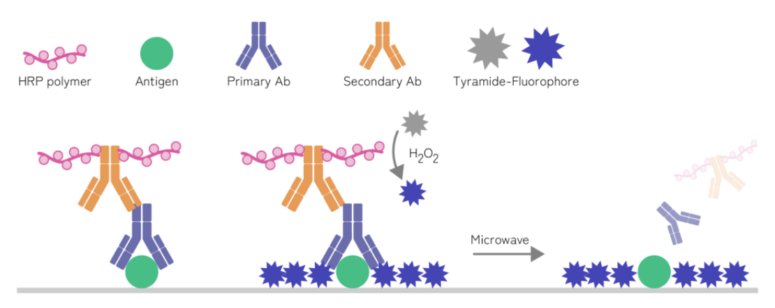
    酪酰胺信号放大技术(Tyramide Signal Amplification, TSA)主要是利用酪胺的过氧化物酶反应。酪胺非活性荧光素底物在HRP和过氧化氢的作用下,会被激活产生活化荧光底物,同时形成共价键结合位点,共价结合在蛋白抗原表面或附近的酪氨酸残基上,抗原和抗体的结合部位就会有大量的酪胺荧光素沉积,使抗原位点处的荧光信号增强。

    酪胺荧光素底物-抗原酪氨酸共价稳定结合,故TSA信号不会受微波影响,可用热修复法清除第一轮与抗原非共价结合的抗体复合物,并能在抗体去除后保留与抗原相关的荧光信号。然后,再用第二种一抗进行第二轮孵育,同时更换另一种酪胺荧光素底物,多次循环反复,不同的酪胺荧光素进行标记就可实现多重免疫组化染色。

    WPS图片(1).png

    产品组成成分

    名称

    AWI0688a

    10T

    AWI0688b

    50T

    AWI0688c

    100T

    保存条件

    TSA-520 荧光染料

    0.5 ml

    2.5 ml

    5 ml

    -20℃,避光

    内源性过氧化物酶阻断剂

    0.5 ml

    2.5 ml

    5 ml

    4℃,避光

    超敏酶标山羊抗小鼠/兔 IgG 聚合物

    0.5 ml

    2.5 ml

    5 ml

    4℃,避光


    保存条件

    荧光染料 -20℃避光保存,12个月

    内源性过氧化物酶阻断剂、超敏酶标山羊抗小鼠/兔 IgG 聚合物 4℃避光保存,12个月


    实验前材料准备

    1、PBS缓冲液、5% BSA-PBS(或者其他封闭液)、柠檬酸钠抗原修复液(或其他修复液);

    2、推荐可用于IHC-P的单克隆抗体;

    3、DAPI染色液(推荐浓度:5μg/mL 货号:AWC0298)


    操作步骤

    1、操作流程简图

    image.png

    2、详细操作步骤

    (1)石蜡切片脱蜡至水:依次将切片放入二甲苯Ⅰ 15min→二甲苯Ⅱ 15min→无水乙醇Ⅰ10min→无水乙醇Ⅱ10min→95%乙醇 5min→85%乙醇 5min→75%乙醇 5min→蒸馏水洗。


    (2)抗原修复:组织切片置于盛满 PH 9.0 EDTA 碱性抗原修复液或者 PH6.0 柠檬酸修复缓冲液的修复盒中于微波炉内进行抗原修复(也可以用高压 1-2min 100℃水煮 15min 95℃水浴 20min 等其他热修复方法)。中火 8min,停火 8min,转中低火 7min,此过程中应防止缓冲液过度蒸发,切勿干片。自然冷却后将玻片置于 PBS(PH7.4)中在脱色摇床上晃动洗涤 3 次,每次 5 min (修复液和修复条件根据组织类型以及抗原类型来确定)。


    (3)阻断内源性过氧化物:切片放入内源性过氧化物酶阻断剂,室温避光孵育 15 min,将玻片置于 PBS(PH7.4)中在脱色摇床上晃动洗涤 3 次,每次 5min。


    (4)BSA 封闭:切片稍甩干后用组化笔在组织周围画圈(防止抗体流走),在圈内滴加用 5% BSA-PBS(或者其他封闭液)均匀覆盖组织,(4)37℃恒温孵育 60  min(此步骤也可以使用山羊血清等封闭液进行封闭)。


    (5)加一抗:轻轻甩掉封闭液,在切片上滴加用抗体稀释液稀释好的的一抗 X,切片平放于避光湿盒内    4℃孵育过夜或者 37℃ 1-2h。(湿盒内加少量水防止抗体蒸发)


    (6)加免疫组化 HRP 二抗:玻片置于 PBS(PH7.4)中在脱色摇床上晃动洗涤 3 次,每次 5min。切片稍甩干后在圈内滴加与一抗相应种属的免疫组化 HRP 二抗覆盖组织,避光室温孵育 30-50min,PBS 洗三次。


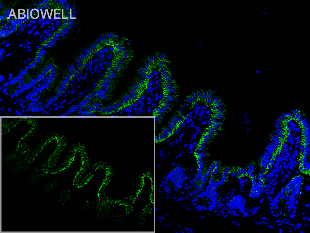
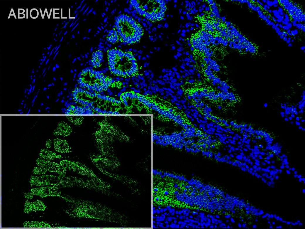
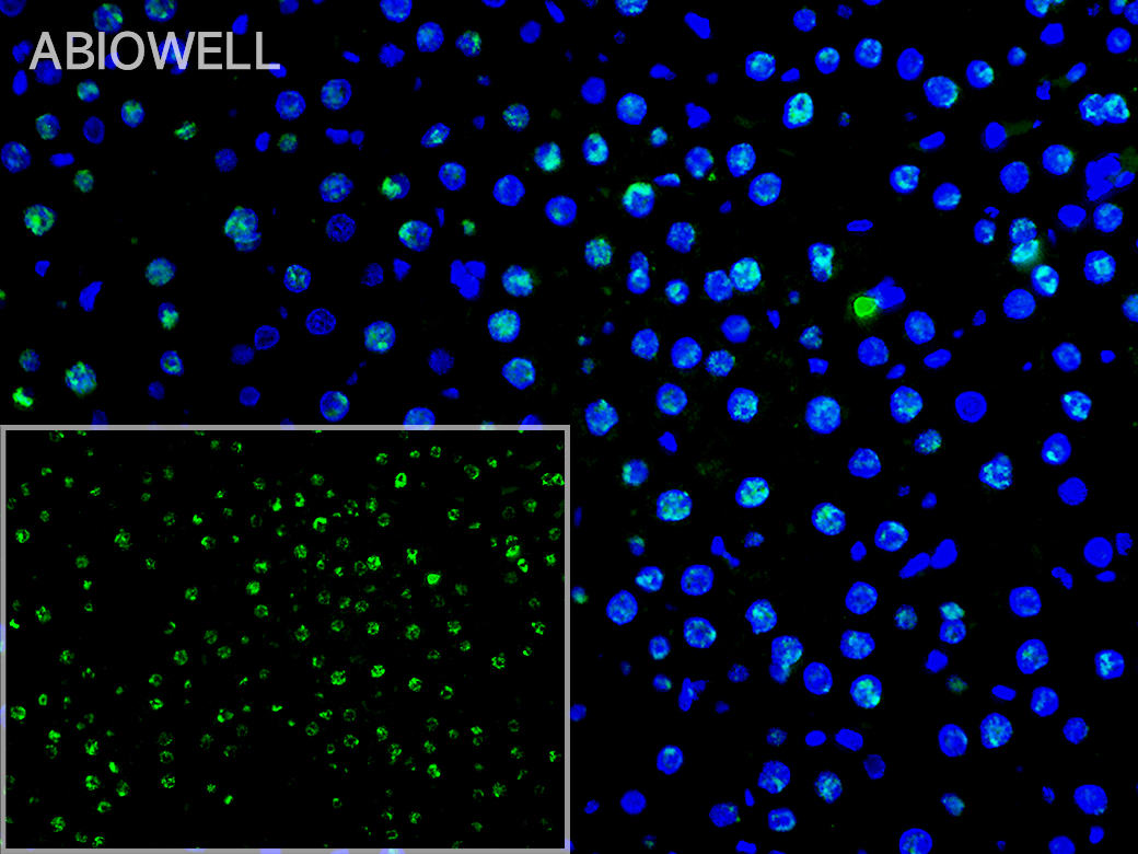
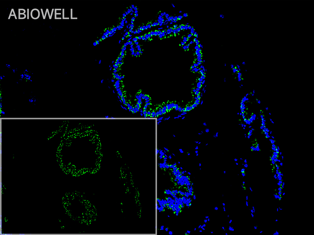
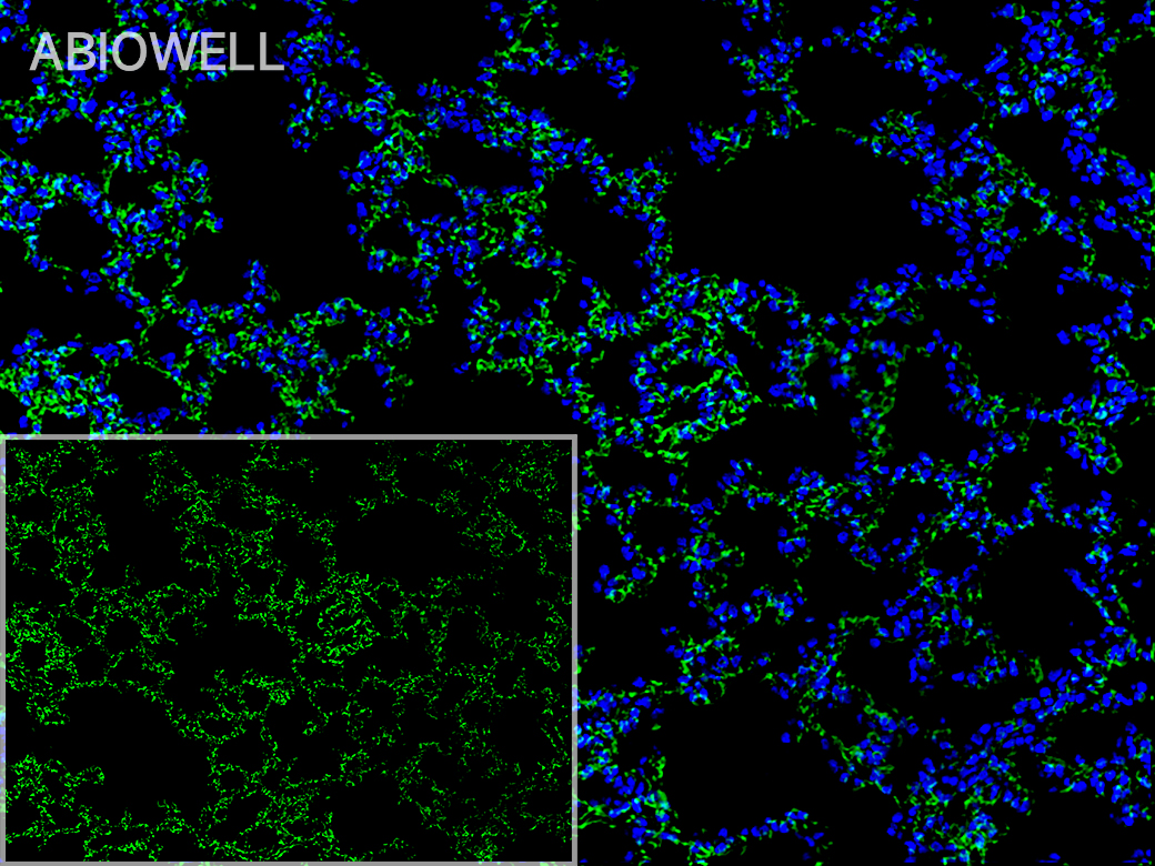
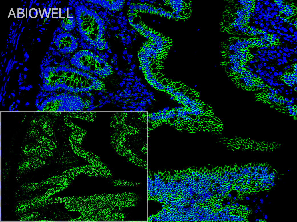
    (7)荧光染料反应:TSA-520 荧光染料反应 10-15 min,PBS 洗三次。


    (8)DAPI 复染细胞核:玻片置于 PBS(PH7.4)中在脱色摇床上晃动洗涤 3 次,每次 5min。切片稍甩干后在圈内滴加 DAPI 染液,避光室温孵育 10min。


    (9)封片:玻片置于 PBS(PH7.4)中在脱色摇床上晃动洗涤 3 次,每次 5min。切片稍甩干后用抗荧光淬灭封片剂封片。


    (10)镜检拍照:切片于荧光显微镜/共聚焦/多通道荧光扫描仪/多光谱成像系统下观察并采集图像。


    染料

    激发波长

    发射波长

    DAPI

    350nm

    420nm

    TSA-480

    450nm

    480nm

    TSA-520

    490nm

    520nm

    TSA-570

    550nm

    570nm

    TSA-620

    590nm

    620nm

    TSA-690

    630nm

    690nm

    TSA-780

    750nm

    780nm


    注意事项

    1、试剂初次使用前请置于4℃解冻,解冻后于4℃短期保存,避免反复冻融,请尽快使用。

    2、为了您的安全和健康,请穿好实验服并佩戴一次性手套和口罩操作。

       3、本产品仅限于专业人员的科学研究用,不得用于临床诊断或治疗,不得用于食品或药品。

    在线客服
    关闭
    小薇 薇薇 小艾 小奥
    联系销售
    技术支持

    image.png

    月琴艾碧维二维码.jpg